TEM

Liquid Phase Electron Microscopy and Spectroscopy of Biological Specimens
Transient, dynamic assemblies of biomolecules in solution are the primary driving forces behind biology. However, studying these at high resolutions requires the use of electron microscopes (EM), which need extremely high vacuums to function.

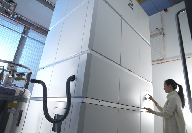
Chromatic Correction
Knoll, the first chromatic aberration-corrected electron microscope in the UK housed at the Franklin, will push the current resolution limits for biological samples by correcting energy variations in the electron beam.

Electron Ptychography and Phase Retrieval in Electron Microscopy
Cryogenic electron ptychography uses coherent diffractive imaging techniques with computational iterative phase retrieval algorithms.

Liquid Phase Electron Microscopy for Cellular Ultrastructure, Biochemistry and Dynamics
Fundamentally, biology is driven by transient, dynamic assemblies of biomolecules in solution.

Electron Diffraction Based Methods of Structure Elucidation
X-ray crystallography has been for many years the primary method of revealing the atomic coordinates of biological and small organic molecules.

Cryogenic electron imaging for investigating the molecular sociology within cells and tissues
Electron imaging under cryogenic conditions allows cells and tissues to be imaged on multiple scales, with pristine preservation of fine molecular detail.

Image Analysis
Many different modalities lead to the generation of large amounts of 2D or 3D image data (TEM, SEM, X-ray CT, LM, etc).

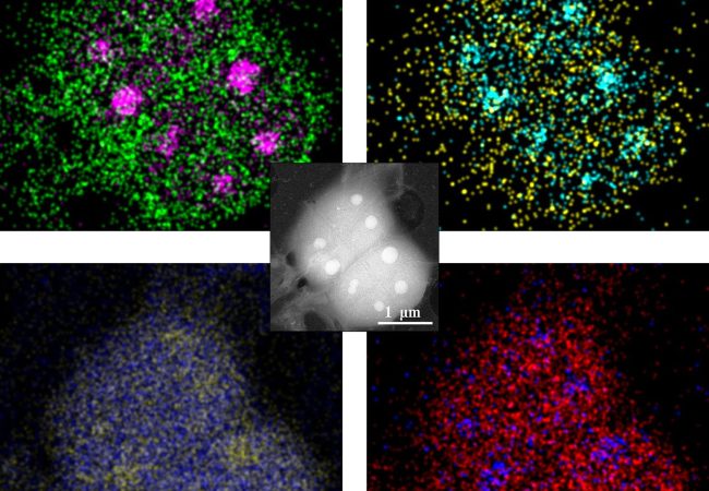
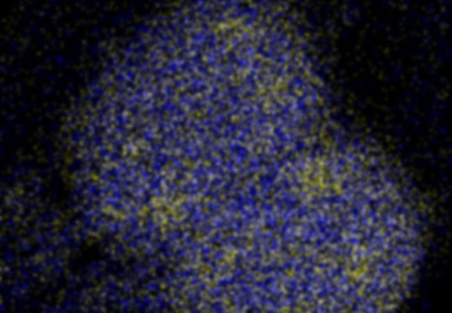
Multidimensional Imaging of Molecular Structures
Our aim: To develop new technologies to see the molecules of life and their dynamics with unprecedented detail.