Dr Michael Grange

Michael completed his D.Phil in structural biology at the University of Oxford, UK, applying in-cell structural biology techniques to investigate the trafficking and egress of viral progeny within cells.
After his doctoral studies, he moved to the Max Planck Institute for Molecular Physiology, Germany, as an EMBO Long-Term Fellow. During his fellowship, he established high-throughput FIB-milling and molecular imaging workflows combining them to investigate the structure and architecture of isolated mammalian muscle and human stem-cell-derived cardiomyocytes. Michael’s group is developing novel imaging approaches to unlock new insight into neurobiology and degeneration, particularly focused on disease initiation and progression.
They use combinations of light and electron microscopy techniques to reveal high-resolution (ultra) structural information at a tissue level in a robust, high-throughput manner. Their goal is to resolve high-resolution structures inside native tissue on a routine basis, transform our understanding of disease at the molecular, cellular and tissue scale and improve disease diagnosis and treatment. The group’s focus is neurodegenerative disorders such as Alzheimer’s disease, imaging the synaptic organisation within the brain during development and building a molecular atlas of how molecular changes occur across different synaptic types.
To do this requires targeting of different synapses to assess molecular content. Additionally, they image how human Tau fibrils impact cellular integrity on a molecular scale, correlating the formation of Tau inclusions with synaptic signals. Molecular observations should unravel new biological and pathological insights. His group works at this interface, demonstrating how new technologies can drive new insight into disease on a molecular scale.
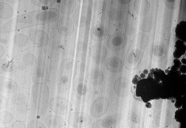
Large Volume Tomography
High resolution large volume tomography with electron microscopy has the potential to transform our understanding of life, by linking the atomic and molecular structure of protein complexes in their biological context – the cell.
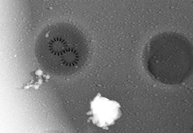
Biochemical Microscopy for imaging across Molecular Scales
Developing a transformative cryogenic 3D biochemical microscope, harnessing the power of high-resolution electron microscopy and mass spectrometry imaging
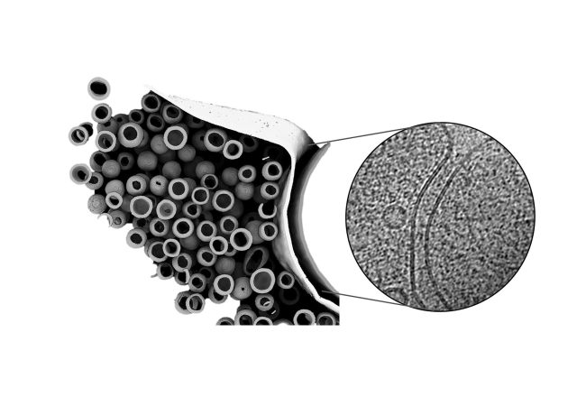
Structural Neurobiology
Developing cutting edge techniques to image structures in the brain on the molecular scale so we can identify early structural changes leading to disease onset.