Dr Jonathan Barnard

Jon is the Electron Microscopy Facilities Manager within Correlated Imaging. Having studied physics at the University of Bristol, Jon studied the structure, chemistry and electronic properties of semiconductors and their defects using state-of-the-art transmission electron microscopes.
After moving to Cambridge University, he played a key role in developing the electron microscope facility at the Department of Materials Science & Metallurgy (MSM). There, he developed 3-dimensional methods for mapping dislocations and chemistry (winning the Japan Institute of Metals medal in 2007) and oversaw the design specification and build of a purpose-built electron microscope laboratory when MSM moved to West Cambridge in 2014, which is now considered to be “one of the quietest buildings in the world”. At the University of York, he developed the York JEOL Nanocentre instrument capability and user base and, played a pivotal role in York’s first cryoEM facility – the Eleanor & Guy Dodson Building (York Structural Biology Laboratory) that opened in 2020.
Jon describes himself as “an evangelical microscopist” and enjoys taking things apart to study in electron microscopes. His move to the Rosalind Franklin Institute marks the next level up in pursuing this line of enquiry – how do living organisms work at the atomic and molecular level? His current interest is in the formation mechanism of bio-composite materials – the nanostructural, crystallographic and chemical hierarchy in teeth and bone.
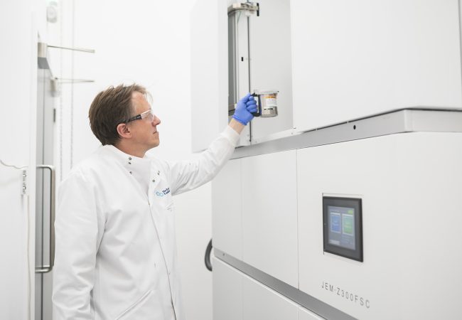
Cryogenic Electron Microscope (cryoARM)
Crewe is a first-generation cryogenic electron microscope based on JEOL’s atomic resolution microscope platform (ARM).
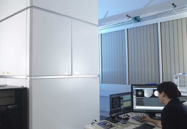
Aberration-corrected transmission electron microscope
Ruska is an aberration-corrected transmission electron microscope (TEM) used to explore novel methods to study radiation sensitive specimens such as biological materials that have been cryogenically preserved or encapsulated in liquid for dynamic observations.
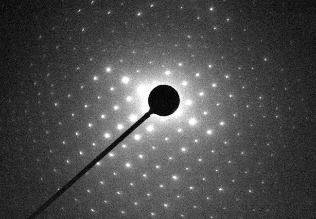
Electron Diffraction
MicroED is an emerging technology that exploits the strong interaction of electrons to reveal the structures of molecules from vanishingly small crystals.
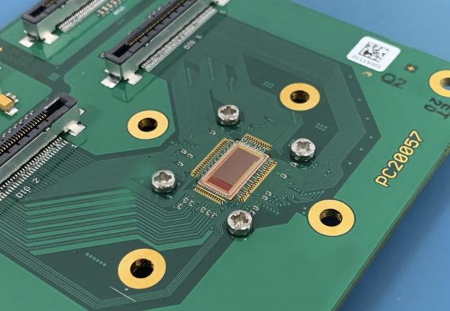
Electron Detector Development
Atomic resolution imaging with electrons causes sample damage. The information per unit of damage is dependent on sample thickness and beam energy.