Professor Angus Kirkland

Angus is Science Director at the Rosalind Franklin Institute, professor materials and Electron Microscopy at University of Oxford and the electron Physical Sciences Imaging Centre at Diamond Light Source.
His research interests include the development and applications of aberration corrected HRTEM for structural studies of nanomaterials, the design of direct electron detectors and electron optics and computational image processing and theory for phase retrieval and quantitative electron microscopy.
Angus completed his MA and PhD at the University of Cambridge using high resolution electron microscopy to study the structures of colloidal metals. Following a post-doctoral Fellowship, he was elected to the Ramsay Memorial Trust Research Fellowship and subsequently as Senior Research Associate in Cambridge.
In 2005 Angus was appointed as professor of materials at Oxford University and in 2011 as JEOL professor of Electron Microscopy. He is the author of over 500 refereed papers and holds 14 patents. He is also Fellow of Linacre College, Oxford.
Angus is the recipient of numerous prestigious accolades, including the 2005 Microscopy Society of America Award for best paper published, the Harald Rose Distinguished Lecture and Prize for Contributions to Image Processing and Exit Wavefunction Reconstruction (awarded in 2015), the Quadrennial Prize of the European Microscopy Society (2016) the RMS Agar Medal in 2017. In 2012 he was appointed as an Honorary Professor, Nelson Mandela Metropolitan University, Republic of South Africa.
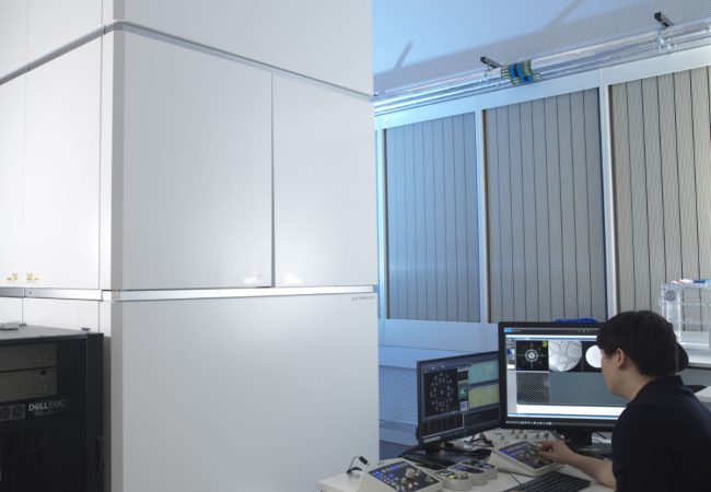
Aberration-corrected transmission electron microscope
Ruska is an aberration-corrected transmission electron microscope (TEM) used to explore novel methods to study radiation sensitive specimens such as biological materials that have been cryogenically preserved or encapsulated in liquid for dynamic observations.
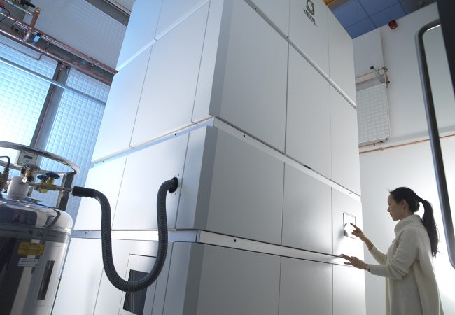
Chromatic Correction
Knoll, the first chromatic aberration-corrected electron microscope in the UK housed at the Franklin, will push the current resolution limits for biological samples by correcting energy variations in the electron beam.
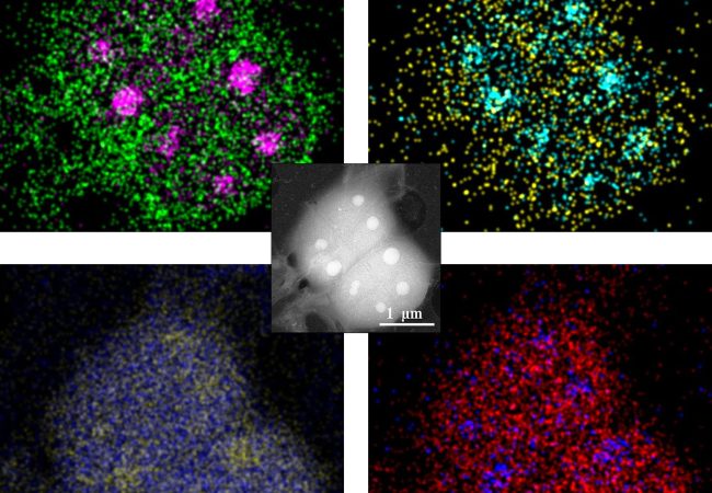
Biochemical Microscopy for imaging across Molecular Scales
Developing a transformative cryogenic 3D biochemical microscope, harnessing the power of high-resolution electron microscopy and mass spectrometry imaging
Cryo-ptycho-tomography
Developing a novel technique using cryo-electron ptychography to perform tomographic characterisation of biological processes at cellular scales, enabling detailed study of rare and complex structures in their native environments.
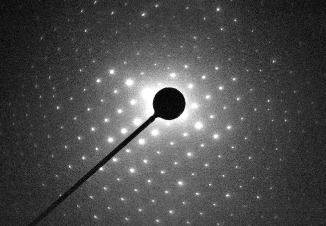
Electron Diffraction
MicroED is an emerging technology that exploits the strong interaction of electrons to reveal the structures of molecules from vanishingly small crystals.
Liquid Phase Electron Microscopy and Spectroscopy
Transient, dynamic assemblies of biomolecules in solution are the primary driving forces behind biology. However, studying these at high resolutions requires the use of electron microscopes (EM), which need extremely high vacuums to function.