Make your own edible cell
Thank you for finding our edible cells page, we are glad you like cells as much as we do!
Here you will find some instructions on how to make your own model cells at home, we hope that you can have fun while learning more about the units of life that make up all organisms.
You will need:
- A cell membrane – a bowl or container
- Cytoplasm – jelly – use gelatine sachets (or suitable alternative) or a packet mix to make this up yourself
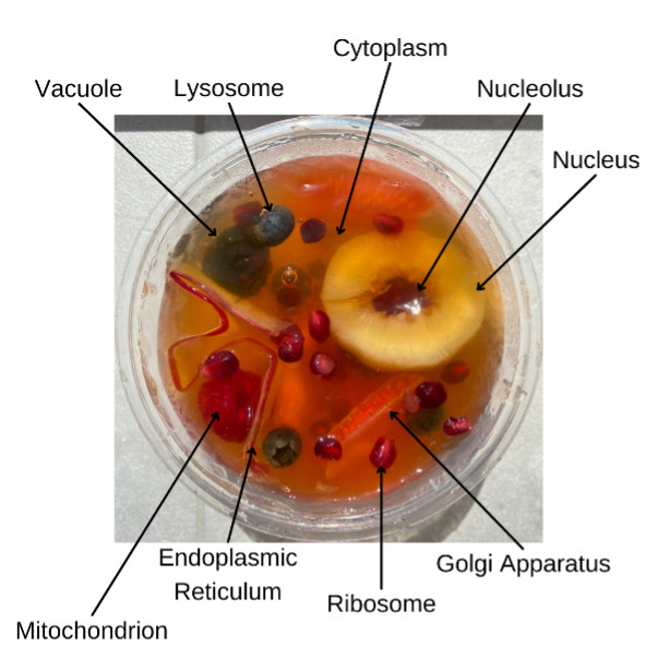
- Organelles of choice – a variety of edible snacks representing your chosen organelles, such as:
- grapes
- slices of banana
- raisins
- nuts
- pitted fruit – for the nucleus and nucleolus
- fruit roll-up
- strawberry laces or gummy worms
- small sweets or chocolates
Note: Using fresh pineapple, kiwi or papaya fruit will prevent your jelly from setting.
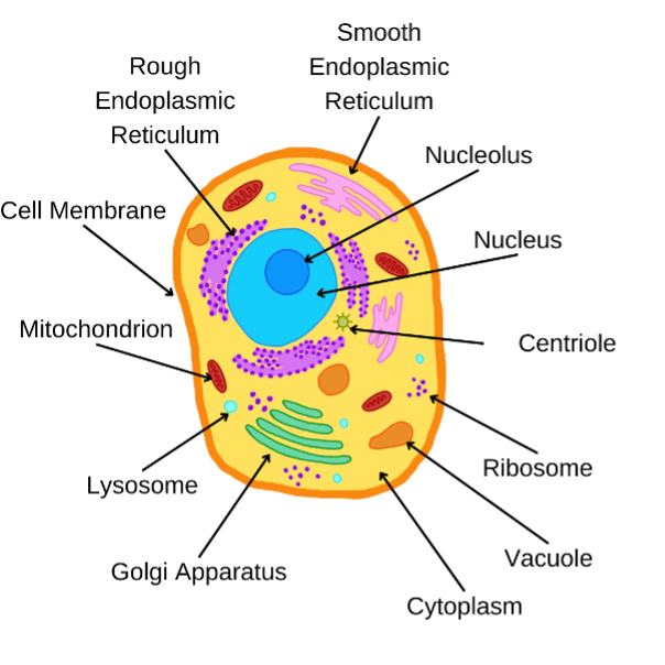
Organelles
Organelles are the subunits in our cells, each one has a specific structure and function. Try to think about what edible snacks you can use that match the structure of the organelles inside our cells.
- Cell membranes provide the shape of our cells and protect the organelles inside, normally these are made from lipids and proteins and they can allow some smaller molecules to enter the cell.
- The cytoplasm is a jelly like substance that is made from water nutrients and waste products from the cell.
- The nucleus is the largest of the organelles and contains all of the cell’s genetic information inside the nucleolus.
- Centrioles are barrel shaped organelles and are usually found in pairs, they help to organise the cell, and are important in cell division
- The endoplasmic reticulum is a continuous membrane that forms multiple flattened sacs, these are involved in the synthesis of proteins and lipids, the rough endoplasmic reticulum is covered in ribosomes
- The golgi apparatus are sac-like organelles that are involved in the secretion and transport of molecules
- Lysosomes are small circular shaped organelles filled with digestive enzymes to help to remove and digest waste or damaged cells
- The mitochondria, often called the powerhouse of the cell, is oval shaped and stores energy for the cell to use
- Ribosomes are small units that float freely or are found embedded in the rough endoplasmic reticulum. Ribosomes are involved in the making of proteins
- Vacuoles are fluid filled organelles that can help to keep the shape of cells, they also help to digest, excrete and store substances
How to make your edible cell
- Make up your jelly mix following the instructions on the packet
- Once your mixture has cooled, pour it into your cell membrane container and set in a refrigerator for 45 mins to 1 hour
- Your jelly should now be soft enough to still insert your organelles but stiff enough that they will suspend
- Start arranging your edible organelles, these can be pushed into the jelly using spoons, straws or even fingers
- Once you are happy with your cell, place it back into the refrigerator to fully set
- Enjoy your edible cell!
Usually to see inside of a cell, researchers have to freeze or fix the cell, like your jelly and only image small slices or sections at a time, this means that you cannot see how organelles move and interact within the cell. Researchers at the Franklin are trying to develop methods to image whole cells and to look at cells in their native liquid states, this would mean that we can investigate these units of life and discover more about how these molecules interact with each other.