BioCOP – the UK’s first multimodular optical microscope – enters testing phase
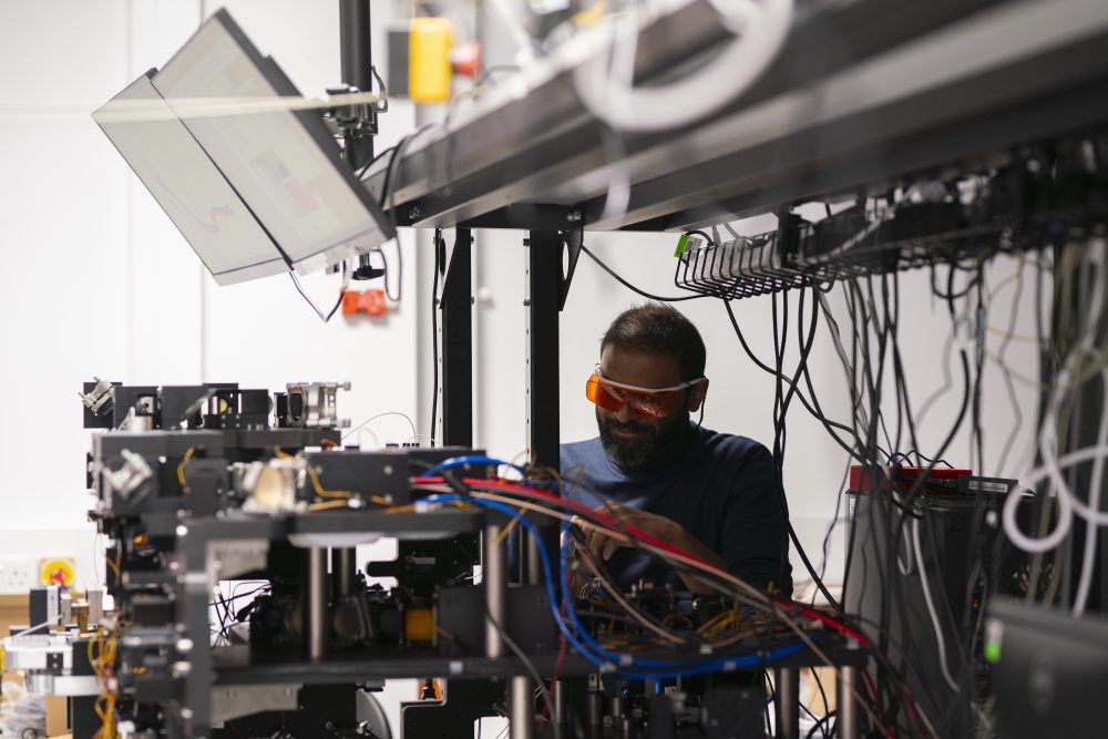
Biophotonic Correlative Optical Platform (BioCOP), which is located at the Rosalind Franklin Institute (the Franklin), has entered its next phase of development of calibration and benchmarking. The instrument is now assembled, and the team will start testing it with model systems and real biological problems.
BioCOP is the UK’s first multimodular optical microscope for high-performance imaging of biological samples across multiple length- and timescales. This versatile platform will enable researchers to study molecular and cellular behaviour within intact and live tissues.
Using this platform, researchers will be able to gain deeper insight into a wide range of biological questions. One of the biological challenges the researchers would like to address is in immunology, to explore the spatiotemporal organisation of the human immune response in health and disease. In host-pathogen interactions, the platform enables real-time, high-resolution imaging of pathogen entry and host-pathogen interactions.
This project is led by Professor Marco Fritzsche, group leader of the Biophysical Immunology Laboratory between the Franklin, and the Kennedy Institute of Rheumatology (KIR) at the University of Oxford, UK. The project represents a strategic synergy between the Franklin and the KIR to bring together technology and instrumentation expertise within the two institutes.
Professor Marco Fritzsche said, “This is an exciting time for the team – we are looking forward to being able to start applying this new technology to biological problems. While developing the instrument, we have been building a network of interested groups, collaborators across our partner institutes, and these groups will be the first to test out BioCOP’s capabilities.”
BioCOP combines minimally invasive Lattice Light Sheet Microscopy (LLSM) and Structured Illumination Microscopy (SIM) based super-resolution imaging in one optical platform. This integration enables large field-of-view imaging of cells in their native states and quantify nanoscale processes such following molecular organisation at unprecedented spatiotemporal resolution.
Compared to a confocal microscope, which is the current gold standard for visualising cell dynamics over space and time, LLSM imaging can produce volumetric data approximately 100 times faster at the same diffraction-limited optical resolution of about 200 nm laterally and 500 nm axially.
This new platform also allows an improved signal-to-noise ratio, enabling clearer and more detailed imaging of biological processes. Rapid deep-tissue imaging with a penetration depth of a few hundred micrometres with subcellular resolution is enabled by the two-photon Bessel-beam light sheet microscopy and an adaptive optics correction system provides a way to compensate for optical aberrations when imaging thick samples. The 3D-SIM technology included in the same platform allows users to switch to super-resolution, achieving double the resolution in all three dimensions.
Dr Narain Karedla, Staff Scientist at the Franklin, explains, “BioCOP works by finely controlling the way light is shaped and directed onto the sample, allowing us to capture incredibly detailed images without disturbing the delicate structures. We use advanced beam shaping to optimize illumination and adaptive optics to automatically correct for distortions caused by thick tissue, a bit like noise-cancelling headphones adjust to block out background noise. This approach enables us to obtain highly sensitive, high-resolution images while keeping the light exposure low to protect live cells during long-term imaging.”
Furthermore, the microscope is equipped with seven excitation lasers and two highly sensitive cameras for correlative imaging. This enables live tracking of cellular protein-protein interactions and organelle dynamics at previously unattainable spatiotemporal resolutions. The added benefit of using lattice light sheet technology is the highly reduced phototoxicity, enabling cells to be imaged for longer without damaging them.
These combined advances mean that the BioCOP platform is uniquely suited for viewing live, dynamic cellular processes in a complex environment with real-time, high-resolution visualisation.
We look forward to keeping you informed about the applications of the BioCOP in the future.