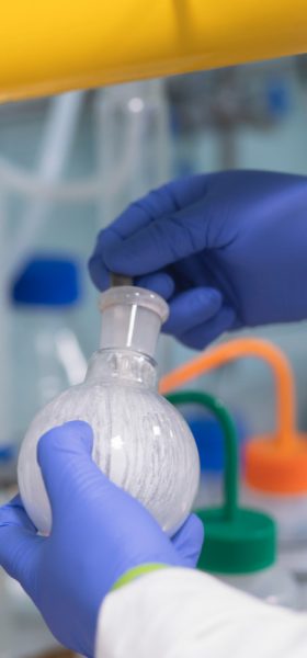
Multidimensional Imaging
Our aim: To develop new technologies to see the molecules of life and their dynamics with unprecedented detail.
“The Franklin is giving us the time and expertise to dream big and create technologies to look more closely at life at a molecular level. We are developing the instruments and techniques which will allow biologists to do experiments they never thought possible.”

The ambition
We will work with industry and academia to create the scientific instruments to revolutionise our ability to image and understand biological molecules. When these machines are complete, we expect the wider scientific community to use them for their research in the future.
Imaging techniques that are being developed now for research may also find applications in the clinic for diagnostic or therapeutic purposes.
What are we doing?
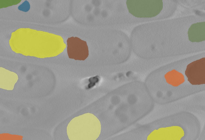
We are developing new technologies that can detect low levels of multiple signals combined with innovative sample preparation.
This will allow us to see biological structures at a molecular level and gain insights into the dynamics of their interactions. We are working to be able to routinely see structures in cellular context with near atomic resolution and to follow how single molecules change over time in performing their functions. The tools to give this information do not yet exist – we are working with manufacturers and developing our own technologies in house to achieve this ambitious goal.
Why?
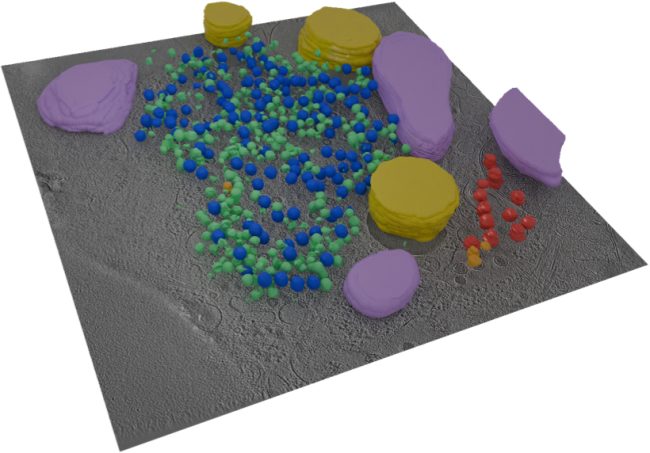
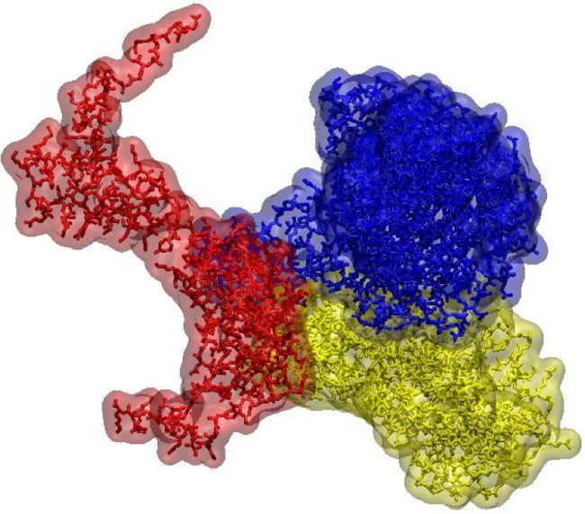
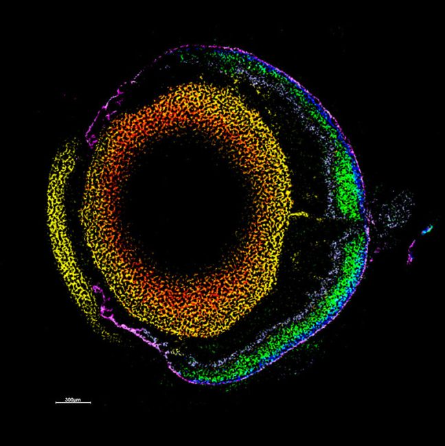
Our work promises new tools that will provide near atomic resolution of molecules in the context of the native cell. Giving valuable new insights into amino acids, proteins, sugars and lipids, including how they function in a healthy system and how their malfunction may contribute to disease. The ability to see interactions between biomolecules is vital as many imaging techniques approaching this resolution are static, whereas in life, biomolecules move and interact constantly.
Research examples
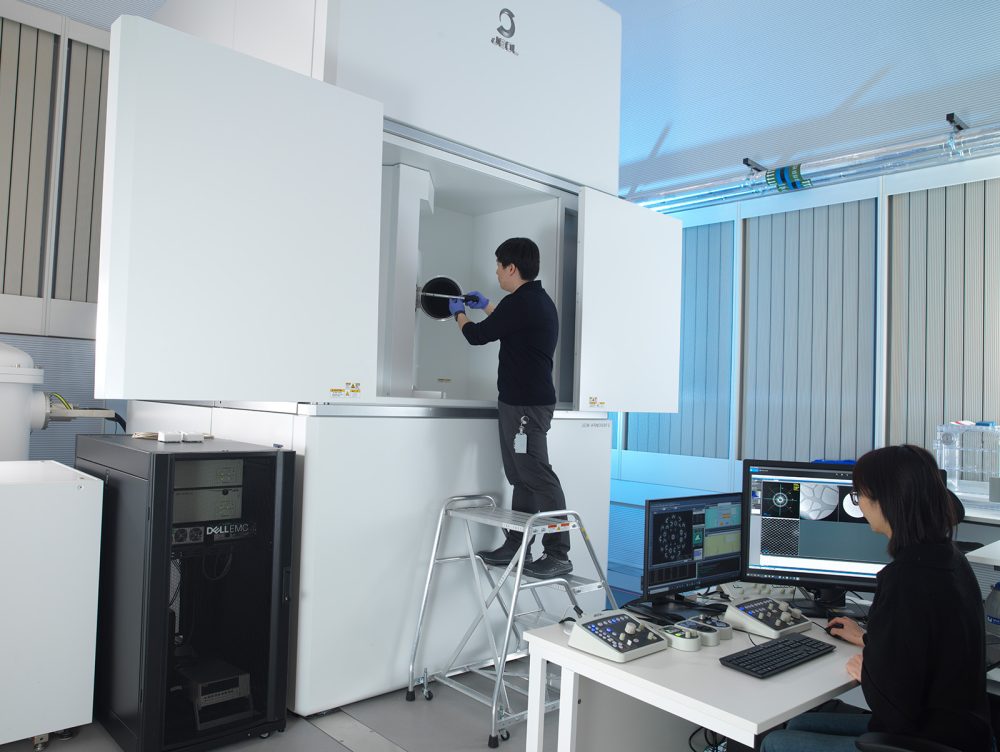
Novel electron microscopy technologies are being developed which could be used to watch how proteins fold and unfold, how molecules and cells interact with their environment, or how a protein changes in response to a drug. We are advancing the microscope hardware, developing detectors, and creating new ways to prepare samples to achieve this.
Why the Rosalind Franklin Institute?
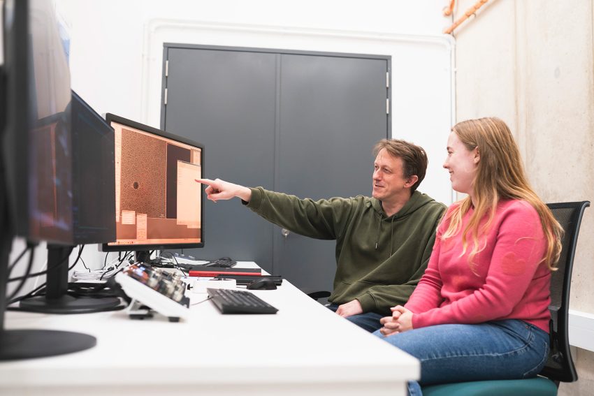
The multidisciplinary nature of the Franklin has allowed us to build a research team with wide ranging expertise including physicists, biologists, chemists, materials scientists and mathematicians. Our outreach into the wider UK research community will allow us to develop tools to meet the needs of a wide range of life scientists.
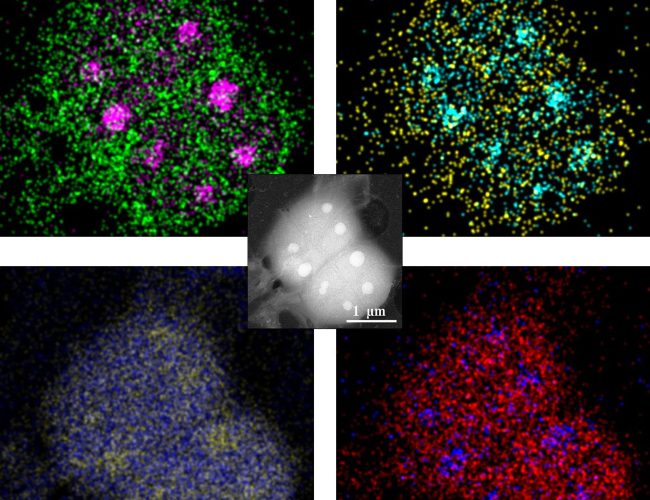
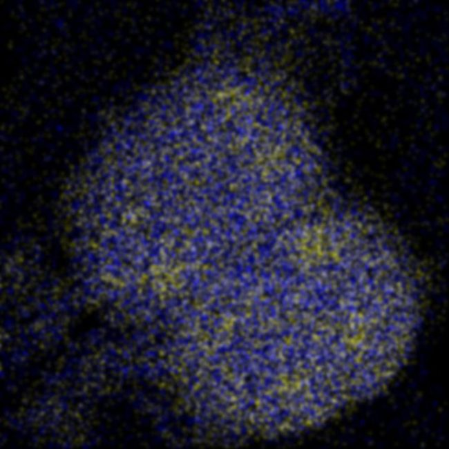
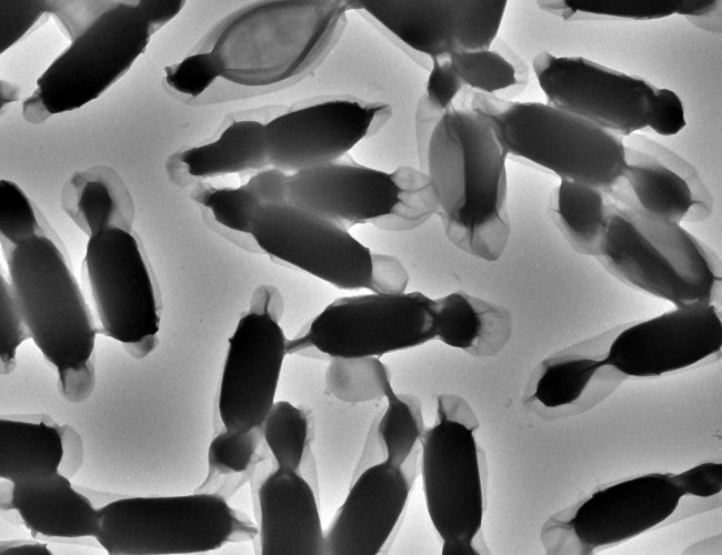
Liquid Phase Electron Microscopy and Spectroscopy
Transient, dynamic assemblies of biomolecules in solution are the primary driving forces behind biology. However, studying these at high resolutions requires the use of electron microscopes (EM), which need extremely high vacuums to function.
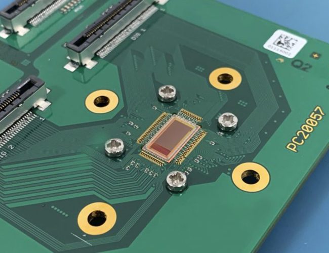
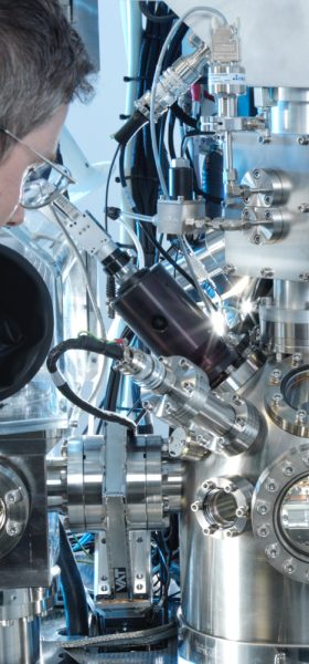
Electron Detector Development
Atomic resolution imaging with electrons causes sample damage. The information per unit of damage is dependent on sample thickness and beam energy.
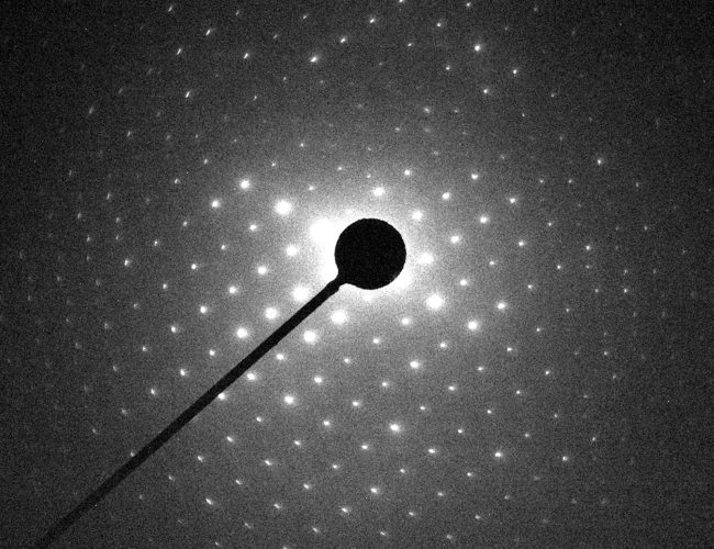
Electron Diffraction
MicroED is an emerging technology that exploits the strong interaction of electrons to reveal the structures of molecules from vanishingly small crystals.
Cryo-ptycho-tomography
Developing a novel technique using cryo-electron ptychography to perform tomographic characterisation of biological processes at cellular scales, enabling detailed study of rare and complex structures in their native environments.
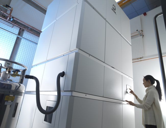
Chromatic Correction
Knoll, the first chromatic aberration-corrected electron microscope in the UK housed at the Franklin, will push the current resolution limits for biological samples by correcting energy variations in the electron beam.