Cryo-ptycho-tomography
Developing a novel technique using cryo-electron ptychography to perform tomographic characterisation of biological processes at cellular scales, enabling detailed study of rare and complex structures in their native environments.
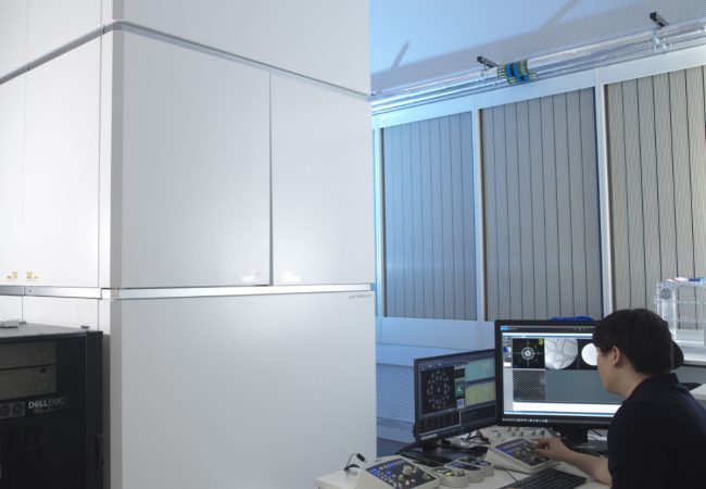
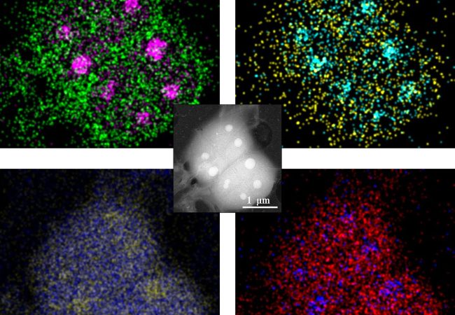
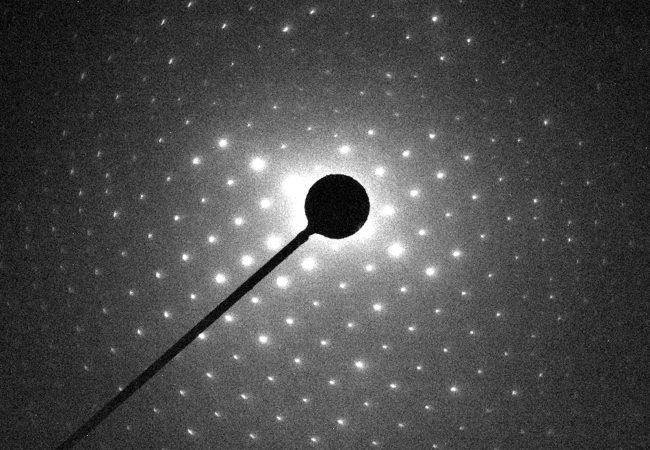
Recent developments in cryo-electron ptychography (cryo-EPty) have demonstrated its potential for characterising biological structures with both high resolution and a large field of view. Key advancements in data acquisition, reconstruction algorithms and instrument automation at the Franklin present new opportunities to extend the capabilities of cryo-EPty to three-dimensional characterisation. At the same time, cryo-electron tomography (cryo-ET) has become one of the most widely used tools in structural biology, enabling the study of complex assemblies in their near-native states.
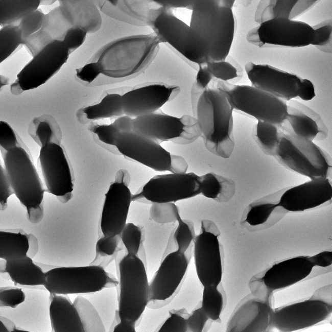
The main scientific objective of this project is to capture three-dimensional, heterogeneous structures of rare occurrence which are most effectively studied within their cellular context. These structures are often challenging to extract or purify for techniques such as single-particle analysis. The increased field of view offered by cryo-EPty compared to traditional cryo-ET is expected to significantly improve the efficiency and success rate of identifying these structures and complexes.
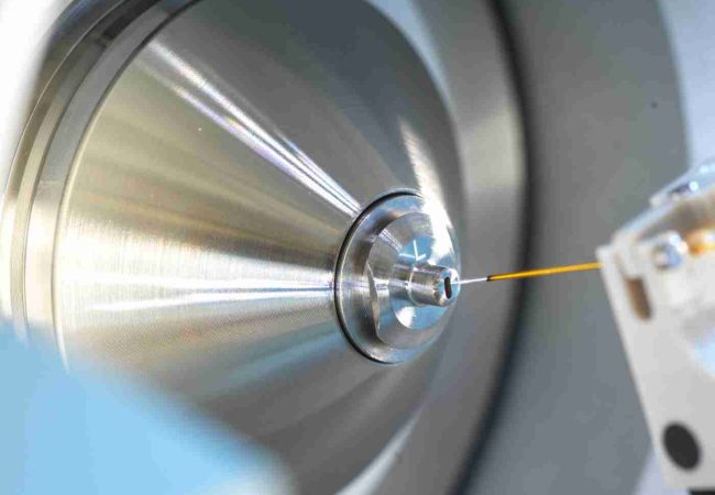
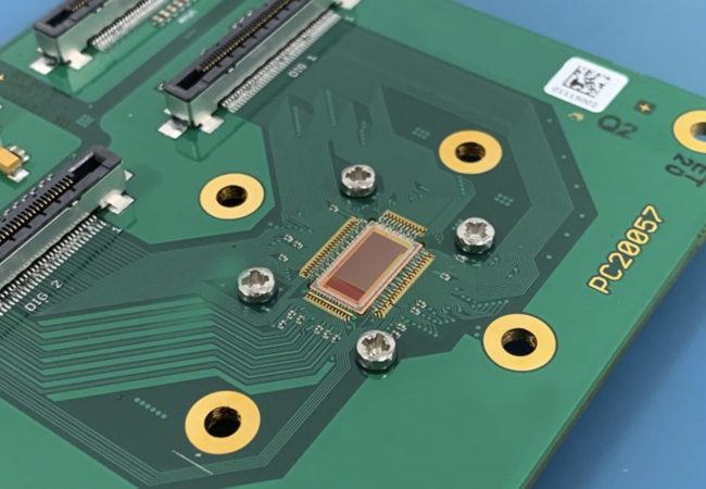
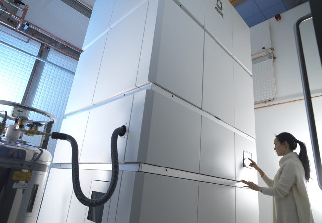
The instrumentation development aspect of the project will integrate several cutting-edge hardware components, in collaboration with our industrial partners, into the established cryo-EPty platform. This integration will pave the way for a novel cryo-electron ptychographic tomography workflow. In addition, the project will also leverage advanced sample preparation workflows available at the Franklin and nearby scientific facilities. This will accelerate progress from the formulation of biological questions to experimental characterisation.