Native ambient mass spectrometry
Native ambient mass spectrometry (NAMS) combines spatial and structural biology by enabling untargeted label-free interrogation of proteins in their functional form directly from their physiological environment.
NAMS has the potential to transform life sciences research by providing unprecedented insight into the transient interactions of proteins with other biomolecules.
Native ambient mass spectrometry enables label-free detection, identification and spatial mapping of native proteins within their natural environment. This provides opportunities for the interrogation of disease progression, drug interactions, and structural and molecular biology more broadly.
At the heart of our NAMS research is the new Thermo Scientific Orbitrap Ascend Structural Biology tribrid mass spectrometer equipped with a nanospray desorption electrospray ionisation (nano-DESI) source built in-house. The mass spectrometer offers high mass range capabilities (up to m/z 16,000). It combines quadrupole, linear ion trap and orbitrap mass analysers allowing multiple rounds of fragmentation (MSn) and opportunities for top-down characterisation of protein complex topology and protein identification in extraordinary detail. The instrument is equipped with a range of orthogonal fragmentation techniques including proton transfer charge reduction (PTCR), electron transfer dissociation (ETD), collision induced dissociation (CID), higher-energy collision dissociation (HCD) and ultraviolet photon dissociation (UVPD).
The instrument offers unique capabilities for NAMS arising from the combination of the bespoke quadrupole, which enables narrow isolation (< 5 m/z) of high mass protein ions, a key requirement for the analysis of complex mixtures of intact proteins and high-confidence separation and assignment of proteoforms which are close in mass, and the high mass range orbitrap analyser which enables analysis of proteins up to 1 MDa.
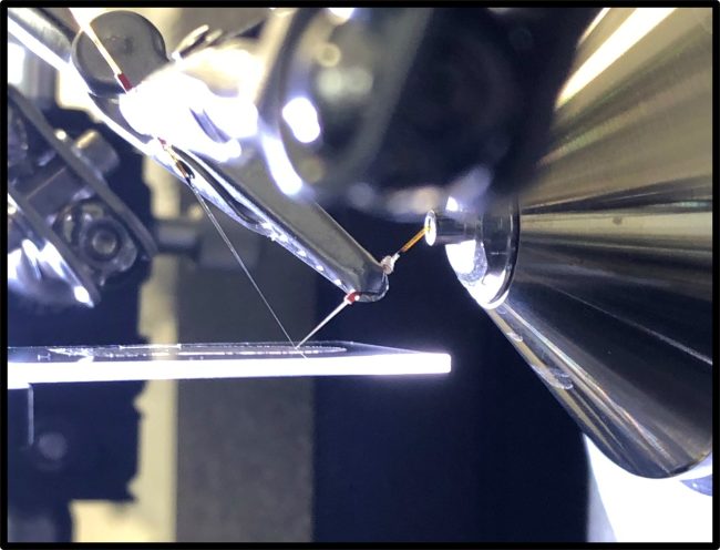
The nano-DESI source enables native ambient mass spectrometry imaging (MSI) of proteins in thin tissue sections with pixel sixes down to 10s of um. Preliminary data taken on the Orbitrap Ascend Structural Biology tribrid mass spectrometer show that protein complexes up to 230 kDa can be mapped directly from tissue.
Collaboration
We are interested in collaborating with those that share our interest in high mass accuracy mass spectrometry.


Thermo Fisher Scientific
Researchers at the Franklin have been working closely with Thermo Fisher Scientific through our Wellcome funded ‘Electrifying Life Sciences’ programme to develop the next generation of electron microscopes for imaging the smallest structures of cells in intact tissues, bring these tools to market and establish new methods and standards for the community to enable their wide use.


Contact information