Mechanistic Proteomics
Proteins are the workhorses of living cells. Proteins operate in a highly dynamic environment interacting with other proteins and other types of molecules including sugars, lipids and nucleic acids.
Fully understanding the identity of proteins and their interactions with their environment reveals the true picture of life.
Both protein expression levels and subtle biochemical changes made to the sequence in the form of post-translational modifications (PTM), dynamically influence biomolecule function. PTM of proteins, for example, is nature’s way of modifying structure and regulating function, interactions, and sub-cellular localisation. The accurate measurement of expression and modification in vivo is essential to understanding the underlying biomolecular determinants of cellular regulation.
We are creating new approaches to characterise protein PTMs on a proteome-wide scale. We are involved in developing new mass spectrometry instrumentation that can accommodate new modes of ion fragmentation including multiple light-, electron- and classical collision- based approaches. This will allow us to characterise PTMs as well as their context (the pertinent protein in its environment). Furthermore, we want to characterise the ion fragmentation chemistry in order to develop automated tools for analysis.
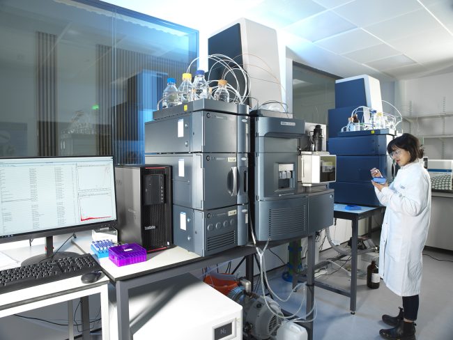
The extension of these methods to other biomolecule classes, such as sugars, allows us to chart new realms of biology. We are improving the sensitivity of cutting-edge experiments with the goal of reaching single cell proteomics. It is little appreciated that the rate and power of mass spectrometry (MS) analysis is outstripping the key chemical techniques that are coupled to it. One of the understated and most powerful aspects of the proteomics experiment is the peptide/protein chromatographic separation: new modes in chromatography have the power unleash the power of MS. We are creating new ultra-high-performance separations for proteomics that are being applied to a range of pertinent biochemicals. One key aspect is our interest in creating ultra-narrow columns which operate at low flow rates (a few nanolitres per minute) to maximise sensitivity.
We are also developing innovative chemical methods for the detection, enrichment and quantification of currently intractable biomolecules as well as difficult protein PTMs such as glycosylation and phosphorylation. These methods operate in several contexts including columns, beads as well as in cellulo.
We apply our approaches to a number of pertinent questions in biology, such as transcription (most recently how the SARS-CoV-2 virus remodels the cell), cell cycle, biomolecule transportation as well as several cell receptors and signalling pathways.


Thermo Fisher Scientific
Researchers at the Franklin have been working closely with Thermo Fisher Scientific through our Wellcome funded ‘Electrifying Life Sciences’ programme to develop the next generation of electron microscopes for imaging the smallest structures of cells in intact tissues, bring these tools to market and establish new methods and standards for the community to enable their wide use.


Contact information