Large Volume Tomography
High resolution large volume tomography with electron microscopy has the potential to transform our understanding of life, by linking the atomic and molecular structure of protein complexes in their biological context – the cell.
Extending and streamlining this capability offers the potential to further our basic understanding of human health.
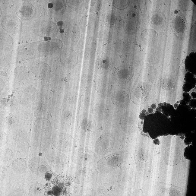
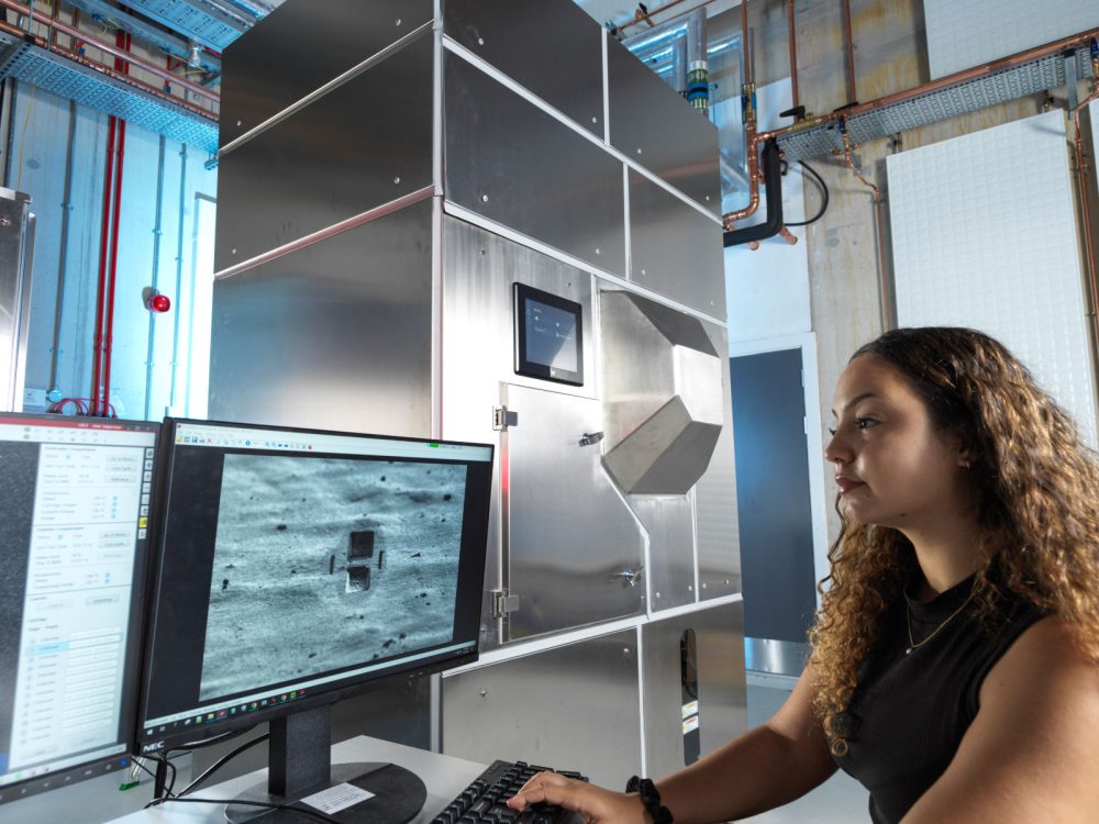
Developing so-called “tomography-in-a-box” – the ability to carry out routine sample shaping for accessing cryo electron tomography (cryo-ET), and extending the volume envelope for productive tomography, is a major focus of the Amplus project. We have been pushing new hardware in collaboration with Thermo Fisher Scientific that allow samples to be shaped using plasma – leading to the development of the first prototype of the “Arctis” Cryo-Plasma-FIB (Cryo-PFIB). The development of this instrument, also in collaboration with Diamond Light Source, enables high throughput lamella fabrication for cryo-ET, leading to the first pseudoatomic structure of the human ribosome in cell (Berger et al. 2023).
Like existing cryo-EM, a technique which has revolutionised our ability to see the molecular structures of life, large volume tomography uses frozen samples, with ice in a glass-like ‘vitreous’ state. These are analysed with an electron beam, giving insight into the atomic and molecular structures of the sample. Using specially prepared samples and analysis, a 3D image of a whole cell or collection of cells could be obtained. Further developments in this project aim to demonstrate a 2-fold increase in the volume attainable with cryo-ET while allowing smaller molecules to be imaged in 3D.
We are looking for partners to develop standardised workflow in both sample preparations, HTP milling and imaging.

Dr Michael Grange
Tomography Group Leader
Dr Maud Dumoux
Deputy Challenge Lead, Quantitative Biology Across Scales

Dr Casper Berger
Senior Application Expert

Dr Tom Glen
Plasma FIB Manager

Dr Matthew Case
Electron Microscope Manager

Dr James Parkhurst
Postdoctoral Research Associate
Dr Calina Glynn
Senior Postdoctoral Research Associate
Dr Gwyndaf Evans
Head of Technology

Dr Jianguo Zhang
Senior Application Expert for in situ Structural Biology

Jake Smith
DPhil Student, University of Oxford
Helena Watson
PhD student

Nadisha Gamage
PhD student

William Bowles
DPhil Student, University of Oxford

Dr Audrey Le Bas
Postdoctoral Research Associate in Biological Sample Preparation

Dr Ravi Teja Ravi
Postdoctoral Research Associate

Thermo Fisher Scientific
Researchers at the Franklin have been working closely with Thermo Fisher Scientific through our Wellcome funded ‘Electrifying Life Sciences’ programme to develop the next generation of electron microscopes for imaging the smallest structures of cells in intact tissues, bring these tools to market and establish new methods and standards for the community to enable their wide use.
Diamond Light Source
As one of the Franklin’s founding members, we have worked closely with Diamond Light Source from our inception. A commissioning call through the Diamond eBIC facility enabled first time community access to key components of our cryo-electron tomography pipeline.

Contact information