High Resolution imaging with secondary ion mass spectrometry (SIMS)
Secondary Ion Mass Spectrometry (SIMS) is a highly sensitive analytical technique offering detailed chemical composition analysis in 3D space with subcellular resolution.

The Franklin is interested in using H2O Gas Cluster Ion Beams (GCIB) to enhance SIMS sensitivity for biological characterisation and achieve multi-omic imaging with sub-cellular resolution.
Time-of-Flight Secondary Ion Mass Spectrometry (ToF-SIMS) is a surface-sensitive mass spectrometry technique widely used for chemical and molecular analysis. The process begins with the formation of a primary ion beam, which then impacts the sample surface. The properties of the primary ion beam are critical, as both its physical and chemical nature significantly influence the outcomes of the analysis. Our system employs two primary ion sources: a Gas Cluster Ion Beam (GCIB) and a C60 ion source.
The GCIB operates with either pure gases such as argon (Ar) or carbon dioxide (CO2), or with water vapor introduced into the ion gun. Large gas clusters, particularly water clusters, offer significant advantages over smaller primary ions like C60. Firstly, large clusters cause less molecular damage to the sample surface, preserving the integrity of the analysed molecules. Secondly, water clusters enhance ionization efficiency, as they contribute additional hydrogens that facilitate the formation of secondary ions, thereby improving overall yields.
Secondary ion formation occurs when the primary ion impact induces sputtering, ejecting material from the sample surface. During this process, a crater is formed, and chemical species are released. Only a fraction of the sputtered material becomes ionised to form secondary ions. The addition of water clusters increases the likelihood of proton transfer, thus enhancing the ionisation process and boosting secondary ion yields.
After sputtering, secondary ions are extracted and directed through the instrument for analysis. The ions pass through a buncher, which time focuses all of the secondary ions, which in turn works to decouple the sputtering process from ion detection. Mass separation occurs in the time-of-flight tube, where a reflectron corrects for initial kinetic energy differences among ions, ensuring improved mass resolution.
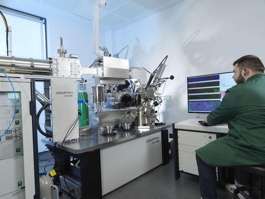
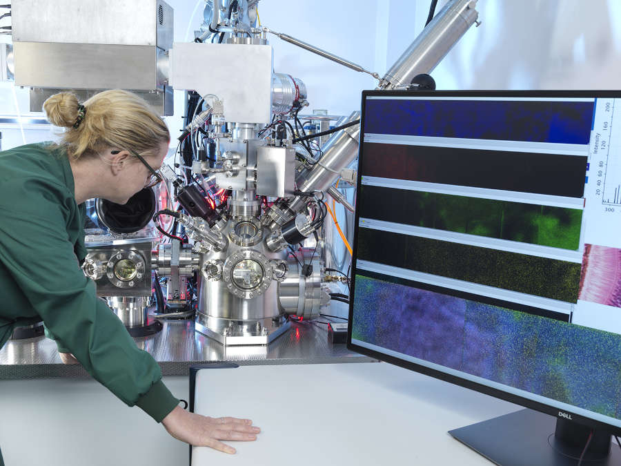
At the Franklin we use the J105 ToF-SIMS instrument developed by Ionoptika Ltd. This instrument offers several advanced features, including continuous-mode primary ion beam operation, cryogenic sample analysis, high-yield detection with water clusters, and the capability to perform both 2D and 3D imaging. Notably, the J105 achieves high spatial resolution, with spot sizes of approximately 1 μm for C60 and 3 μm for GCIB, surpassing the resolutions of many commercially available systems that typically operate at tens to hundreds of microns.
Our research group is actively working on further advancing the capabilities of the J105. One key focus is improving spatial resolution through the integration of an electron cyclotron resonance (ECR) plasma source. This upgrade aims to reduce the spot size to a few hundred nanometers, enabling detailed cellular and subcellular analyses. Additionally, we are developing laser post-ionisation techniques to significantly increase secondary ion formation, thereby minimising signal loss.
We are currently engaged in collaborations with a diverse range of research groups. Within the Rosalind Franklin Institute , we are working with Dr. Siva Ramadurai on imaging gold nanoparticles in HeLa cells, with Dr. Maud Dumoux and Dr. Michael Grange’s groups on correlative SEM-SIMS imaging, and with Dr. Brian Caffrey on assessing chemical damage in bacteria, among other projects.
Externally, we are collaborating with research groups on several initiatives, such as analysing protein fragmentation and ionization (University of Manchester), developing microscope-mode SIMS (University of Oxford), and imaging lipofuscin particles in mouse brains (Yale University), as well as others.
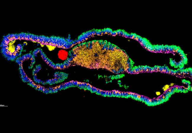
Our instrument is highly versatile, capable of analysing a wide variety of surfaces of interest, with a special resolution of between 1 μm (C60) and 5 μm (H2O clusters). We can generate both 2D and 3D chemical images, allowing us to investigate the spatial distribution of chemical species across the surface and throughout the depth of the sample.
Furthermore, we are exploring methodologies for correlative imaging, combining ToF-SIMS data with scanning electron microscopy (SEM). This integrative approach will enhance our ability to analyse and interpret complex cellular and molecular information, offering new insights into biological systems. Together, these advancements aim to establish SIMS as a state-of-the-art tool for high-resolution, high-yield mass spectrometry imaging.


Ionoptika Ltd
The instrument developed in collaboration with UK based Ionoptika enables a dramatic improvement in mass spectrometry imaging (MSI). The new instrument is capable of imaging the whole surface simultaneously, allowing more accurate imaging at faster speeds than was previously possible.


Contact information