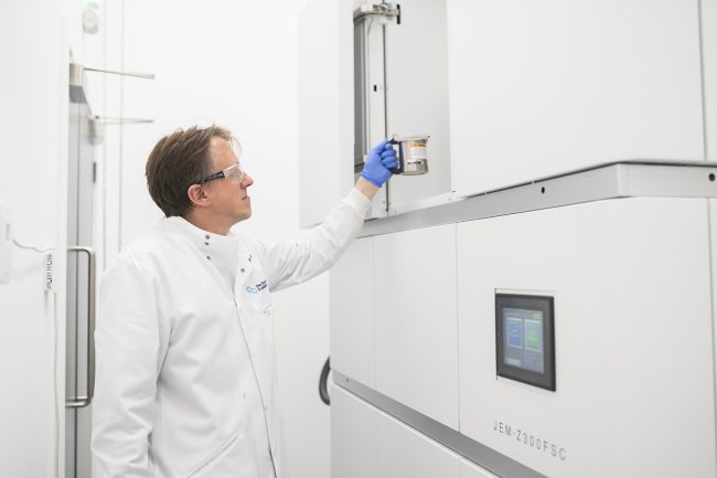
Cryogenic Electron Microscope (cryoARM)
Crewe is a first-generation cryogenic electron microscope based on JEOL’s atomic resolution microscope platform (ARM).
The high brightness cold-field emission gun, in-column energy filter and robotic stage allows detector agnostic imaging and diffraction with or without energy filtering. This provides a platform to test state-of-the-art candidate detectors for biological electron microscope applications.
Crewe is a cryogenic transmission electron microscope based on JEOL ARM platform (first generation cryoARM). It represents the state-of-the-art ‘work horse’ instrument used to determine protein structures all around the world.
Here at the Franklin, we use Crewe to test new techniques based on imaging and diffraction experiments to extract the maximum amount information encoded by each electron passing through the (biological) sample. This involves adapting detectors, often developed by the high-energy physics, e.g. at the large hadron collider (LHC), where radiation tolerance and high-fidelity measurement is critical to understand the nature of our universe.
Crewe is operated at a range of energies (100, 200 and 300 keV) and its illumination system is capable to varying the electron current over six orders of magnitude (tens of femto-amps to tens of nano-amps) with a broad range of beam size and angular dispersion. This is all due to the high brightness cold field emission gun sitting at the top of the column.
The in-column energy filter (Omega type B) can select a range of scattered electron energies with high acceptance (large field of view or large scattering angle) without affecting the camera/detector choice in the camera chamber. This allows us to quickly retrofit candidate cameras and sensors, often optimized for specific electron beam energies or applications.
Finally, the cryogenic robotic stage and automatic loading system (cryoSPECPORTER) allows up to twelve samples to be studied, often using automatic routines, e.g. single particle analysis (SPA), electron tomography or micro-electron diffraction (μED), with its high tilt range (±75o) and optically encoded stage positioning system. This allows us to flexible program the microscope to automatically find and study regions of interest through Python scripting or third-party applications, e.g. SerialEM.
Crewe provides a of state-of-the-art system and that can be adapted and customised quickly, providing a platform to testing new ideas through hardware, scripting or serendipitous tinkering.
Operating energies: 100, 200 and 300 keV
Gun type: tungsten <310> cold field emission
Condenser optics: 4 lenses with eight (8) apertures: 150, 100, 70, 50 (CLA1) and 40, 20, 10 and 5 μm (CLA2).
Number of samples: 12
Sample tilt range: ±75o
Operating temperatures: 80 K (cryo) or 300K (room temperature)
Detector mounting: below view screen, stackable block system, up to five detectors.
Current detector configuration: Gatan K2 (300 kV imaging), Timepix4 (200 and 100 kV diffraction).

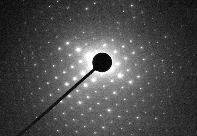
Electron Diffraction
MicroED is an emerging technology that exploits the strong interaction of electrons to reveal the structures of molecules from vanishingly small crystals.
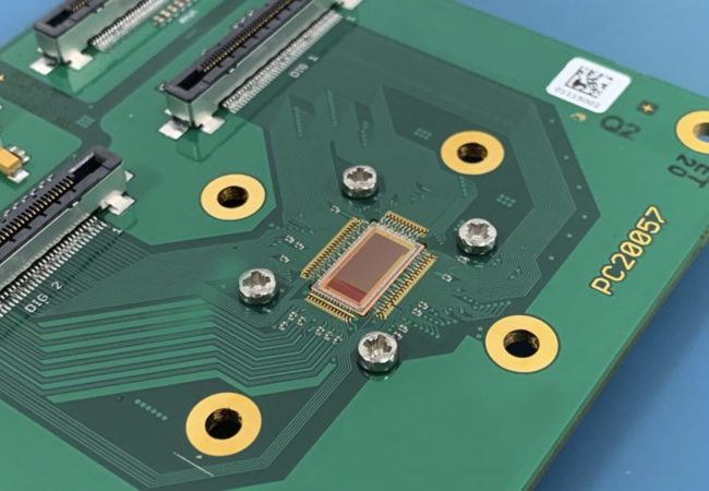
Electron Detector Development
Atomic resolution imaging with electrons causes sample damage. The information per unit of damage is dependent on sample thickness and beam energy.
Contact information