Transmission electron microscopy (TEM) of frozen samples (Cryo-TEM) is used to examine biological samples in fine details and relies heavily on the formation of vitreous (glass-like) ice. Sample preparation, including the format of ice within the frozen samples, is crucial to the quality of data generated in subsequent TEM analysis.
The production of the required ice structure for high quality data generation can be a time-consuming and challenging process. It does not always yield ice which is optimal for cryo-TEM analysis. Typically, samples must be handled in specialist low-humidity rooms, with scientists wearing masks and filtering liquid nitrogen to minimise any risk of contamination. Other options include commercially available robotic systems which can help to automate the freezing process for sample preparation.
About the innovation
Our researchers have discovered a new and reproducible way to generate high quality vitrified biological samples for TEM. This is achieved by creating graphene liquid cells, comprising a layer of graphene below and above a layer of liquid sample suspended in a gold grid.
This configuration does not require pre-freezing, which can remove a lot of the challenges and steps which typically introduce crystalline ice contamination. Graphene liquid cells can be placed directly into the TEM in cryo mode (80K), which then freezes in situ due to liquid nitrogen temperatures and high vacuum to reproducibly form vitreous ice.
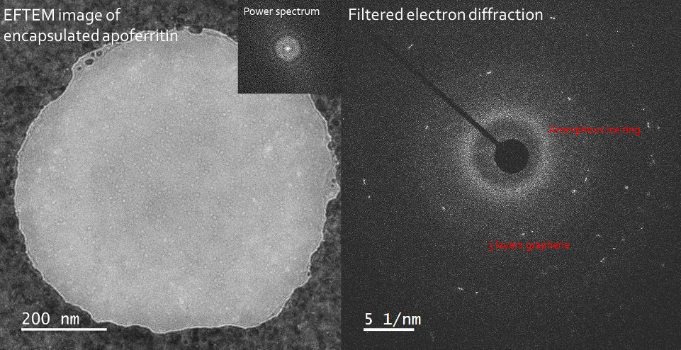
Example image demonstrating the quality and uniformity of ice produced by this method
Advantages
Reproducible, high-quality vitreous ice for TEM.
Removes the need to handle vitreous ice outside of the microscope, reducing the risk of contamination.
Compatible with samples as thin as 30nm.
Generates a uniform thickness sample, meaning 100% of grid area is usable.
Applications
Sample preparation for cryo-TEM
Publications: TBC
Patent Status: PCT filed
Opportunity: Commercial license or co-development.