Proteins naturally present in biological samples are extremely important in medical research and their presence, absence or dysregulation can act as a marker of disease. Broadly, two approaches exist for analysis by mass spectrometry. ‘Top-down’ aims to analyse native proteins whole whilst ‘Bottom-up’ typically uses chemical or enzymatic digestions to break bonds and generate smaller peptides for mass spectrometry analysis.
Mass spectrometry imaging (MSI) – incorporating techniques such as MALDI, DESI and LD-REIMS – can provide powerful, untargeted insight into the chemical makeup of biological tissues and cells. In MSI, both “top-down” and “bottom-up” approaches can be limited by several drawbacks. The dynamic range of the proteome can limit top-down proteomic analysis, where multiple different proteins of vastly differing mass and concentration are present. In contrast, the use of liquid-phase enzyme solutions to digest samples in bottom-up approaches can delocalise analytes, bottlenecking image resolution.
Approaches which can digest samples without enzymatic/liquid reagents can conserve the resolution of typical MSI, whilst overcoming the difficulties in dynamic range typically associated with top-down proteomics.
About the innovation
The invention uses electrical current to generate a cold argon plasma, which can then be directed through a needle to generate a corona discharge. This discharge can be used to digest condensed phase samples for subsequent analysis using MSI. Digestion products include ‘classical’ abc-xyz peptide fragments, with a proposed mechanism that is more heavily reliant on free-electron or radical mediated reactions.
This invention has been applied to detect the presence/absence of a known protein in a cell lysate sample, with good success. We have also been able to detect protein which has been spiked on to biological tissue samples in a selective manner, depending on the exposure to our argon plasma digestion.
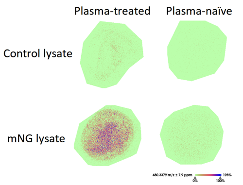
Figure 1 : MALDI-MSI heatmap of a peak at m/z 480.34 in lysates from control cells and cells modified to express the fluorescent protein mNeonGreen, where the lysate is either plasma-naïve or has been exposed to the cold argon plasma setup for one minute. This ion is present only above noise in the plasma-exposed cell lysate where mNeonGreen is present.
In addition to the spiked tissue samples, cell lysates known to express/not express a protein (mNeonGreen), have been proven to be selectively detected using the invention.
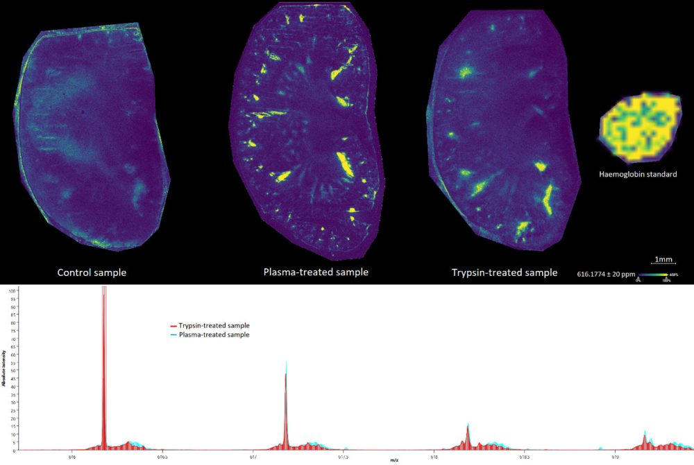
Figure 2 : MALDI-MSI heatmap of the peak at 616.18 m/z (haemoglobin) across kidney tissue samples either untreated, plasma treated, or trypsin treated. Spatial resolution is visibly improved with plasma treatment, allowing for observation in both inner and outer medulla. Plasma treatment has comparable peak intensities compared to that of trypsin treatment.
Advantages
Fast – digestion takes minutes compared to typical overnight digestion with trypsin.
Simplified sample preparation.
Condensed phase digestion – doesn’t require samples to be in the liquid phase.
Enhanced spatial resolution – analytes are not delocalized using APD, so spatial resolution in MSI is maintained.
Can be built into automated workflows for sample digestion.