Using artificial intelligence to understand stillbirth
Researchers in the Rosalind Franklin Institute’s Artificial Intelligence (AI) and Informatics theme are collaborating on a bold international programme to find ways to reduce stillbirth rates by half in just three years.
As one of the Franklin’s founding members, we have worked closely with Diamond Light Source from our inception. A commissioning call through the Diamond eBIC facility enabled first time community access to key components of our cryo-electron tomography pipeline.


Somewhere in the world, a baby is stillborn every 16 seconds. With 2 million stillbirths globally each year, advances in how gestational development is measured, modelled, and predicted could help clinicians to better recognise and prevent stillbirth.
Having a measurable impact on a global problem like stillbirth requires bold approaches to science, with teams coming together from across lots of different disciplines.
A global initiative to reduce stillbirth rates
AI and informatics researchers at the Franklin play a critical role in the Wellcome Leap In Utero programme. This $50 million international initiative aims to rapidly develop and apply new understanding about how complications in pregnancy might lead to stillbirth.
Bringing together X-ray imaging at the Diamond Light Source’s Synchrotron facility with AI and machine learning capabilities at the Franklin, the AI and Informatics team is tackling the challenge by developing rapid new ways to analyse images of placenta tissue samples.
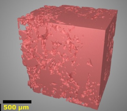
In this part of the In Utero programme, after babies are delivered, samples of placental tissue from the mothers are collected, prepared and annotated for imaging by collaborators at the Universities of Manchester and Birmingham. The 1.5mm tissue samples are then sent to Diamond Light Source, where they are imaged using very concentrated, high-energy X-rays. Here, many 2D images are generated by rotating the sample, which are then reconstructed into large, highly detailed 3D images.
Automation at the Franklin
The images are passed on to the Franklin, where the team uses their advanced image analysis software to analyse key structures in placental tissue using a process called segmentation. Powered by machine learning and AI, the software classifies each pixel in the 3D image into either maternal or fetal blood vessel or tissue. e. It then extracts relevant geometric and ultrastructural information.
Data from the Franklin’s detailed segmentation analysis is then passed up the chain to other In Utero programme collaborators where it is used to power simulations and predictive models. These help to understand how different placental tissue characteristics might cause stillbirth.
“The work we do at the Franklin is one step in a long process that brings together a whole host of global collaborators.”
“The work we do at the Franklin is one step in a long process that brings together a whole host of global collaborators,” says Dr Michele Darrow, the Franklin’s Head of Data Strategy and Cryo-Electron Imaging. “It is a challenge – our work represents a step-change in the amount of data available for downstream analyses. By working collaboratively in this way, the Franklin is contributing to a complete analysis of the role of the placenta, helping to relate complications in pregnancy to the structure of the placenta.”
Scaling up image analysis
For Dr Darrow, taking part in the In Utero programme presented an exciting opportunity. The team needed to find ways to improve, scale and automate existing software so it could rapidly extract useful data from hundreds of 3D images of placental tissue.
Dr Darrow explains: “Conventionally, image analysis involves literally tracing every organelle, tissue or vessel by hand. But this isn’t tenable when you’re looking at more than a handful of datasets – it creates a bottleneck. So we have to train AI to automatically recognise and segment different biological structures in each image. We have a small, effective computer model from a previous study on healthy tissue, so our challenge has been to adapt this with new techniques and methods so we can look at unhealthy tissue.”
The segmentation software used in this project was developed over the past four years by Dr Avery Pennington, Senior Research Software Engineer, who joined the Franklin from Diamond Light Source. Dr Pennington works to scale up the machine learning model by training it with knowledge and insights from other disciplines.
“One of our biggest hurdles is to train the segmentation software so it can analyse images far more rapidly,” says Dr Pennington. “To do this, we’re incorporating very niche knowledge about the microscopic structures of a placenta that typically only a pathologist would know. It’s challenging but very exciting to work across lots of disciplines in this way.”
Developing the Franklin’s capabilities in automated image analysis will have a far wider reach than the In Utero programme. The team is committed to making their software, models and data open source, so other research teams around the world can apply the enhanced tools developed.
Dr Pennington says: “We are developing a variety of new imaging methods for the In Utero programme. A lot of these could be applied to other types of synchrotron data, so our software tools can be used to help answer many other important questions about human health.”