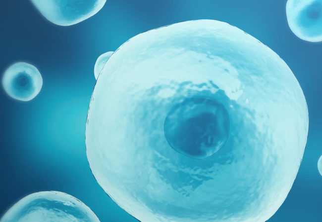
Synthetic biology solutions for imaging
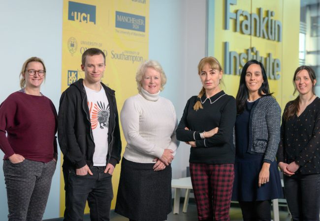
Five EPSRC Impact Acceleration Account funded projects strengthen the ties between the Bristol BioDesign Institute and the Rosalind Franklin Institute.
Five EPSRC Impact Acceleration Account funded projects strengthen the ties between the Bristol BioDesign Institute and the Rosalind Franklin Institute.
In 2022, the Bristol BioDesign Institute (BBI) at The University of Bristol was awarded EPSRC Impact Acceleration Account (IAA) funding to develop links with The Rosalind Franklin Institute. The aim of these awards is to support knowledge exchange and impact from EPSRCfunded research. One way to achieve this is by fostering collaborations between research organisations.
‘Our research portfolio has obvious synergies with The Rosalind Franklin Institute and we already had various long-running pairwise collaborations with colleagues there; this award allowed us to explore further opportunities to bring together the expertise and research facilities of the two institutes and deliver impactful research,’ explains Kathleen Sedgley, Scientific Manager of the BBI.
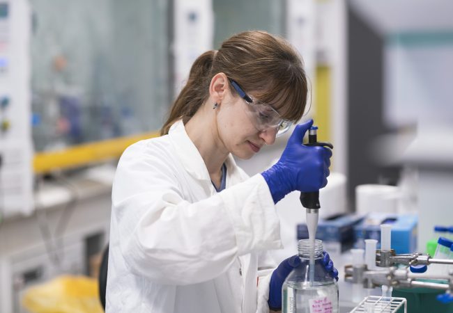
The BBI issued a call for research proposals involving scientists from the BBI and The Rosalind Franklin and funded five short-term projects that ran from February until the end of June 2022. The overarching goal of these projects was to develop synthetic biology solutions for imaging molecules in living cells.
Professor James Naismith’s team has been working with Professor Dek Woolfson, Dr Mark Dodding and Professor Paul Verkade at the BBI on variations of a de novo synthetic peptide system1 that can enter mammalian cells and label specific proteins for correlated light and electron microscopy.
‘The major challenge in transmission electron imaging of human cells is the inherently low contrast, which makes it hard to distinguish proteins from each other,’ says Professor Naismith. ‘The possibility of delivering peptides that carry fluorescent and electron-dense cargoes to subcellular targets will help locate structures of interest specifically and precisely,’ he adds.
So far, they have demonstrated by light microscopy that they can dual-label proteins with both fluorophores and heavy atoms. Their next steps will be to image the cells with an electron microscope and, if they are successfully labelled, apply for a larger grant to continue developing this game-changing technology.
Dr Marco Fritzsche’s group has teamed up with Professor Christoph Wülfing at the BBI to explore how mechanical cues within the tumour microenvironment may influence the ability of cytotoxic T lymphocytes (CTLs) to kill tumour cells. They are using bio-synthetic mechanosensors2,3 to quantify the biomechanical force exerted on specific receptors on CTLs co-cultured with tumour cells in in 2D and 3D systems. This project has the potential to transform cancer immunotherapy by offering new insights into the effects of the microenvironment on tumour immunosuppression.
Bringing together the BBI’s expertise in determining cytotoxic T cell function in 3D tumour cell models and the Rosalind Franklin’s microscopy approaches to measure forces on cells in real time is proving to be very fruitful

With the IAA funding they have been able to show that cells in 3D culture systems can be labelled with beads and that bead displacement can be used to measure biomechanical forces.
Another project involves a collaboration between Dr Michael Grange, Professor Mark Dodding and Dr Paul Verkade and aims to integrate two imaging technologies — focussed ion beam (FIB) milling and in situ cryo-electron tomography — to examine the inside of microtubules with unprecedented resolution. The presence of filamentous actin in the microtubule lumen4 suggests that further investigation into this subcellular compartment could offer new insights into microtubule dynamics and functions.
Professor Ray Owens and Professor Imre Berger are examining cryo-EM structures of the G-protein-coupled receptor FFAR1, which has been implicated in long COVID and type 2 diabetes. They are assembling ‘megabodies’5 to stabilise the receptor coupled with water soluble inhibitors. Understanding how such inhibitors modulate the activity of FFAR1 will offer clues for developing new therapeutics for both these conditions.
‘We are aiming to complete the work started under this collaboration through an award from BrisEngBio, the BBI’s new Centre for Engineering Biology, with a view to obtaining longer term funding for the project,’ Berger explains.
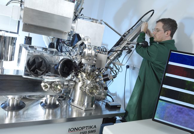
Last but not least, Dr Mark Basham and colleagues are using the IAA seed-funding to speed up the processing of cryo-EM data using novel cluster technology (DisTRaC).
Commenting on the overall experience, Professor Naismith says: ‘With IAA funding we’ve been able to pursue a broad range of high-risk projects that fit with the Institute’s vision and strengthen our ties with the BBI,’ Naismith says. ‘It’s been a win-win.’
References: 1. Rhys GG, Cross JA, Dawson WM, Thompson HF, Shanmugaratnam S, Savery NJ, Dodding MP, Höcker B, Woolfson DN. De novo designed peptides for cellular delivery and subcellular localisation. Nat Chem Biol. 2022 Jul 14. doi: 10.1038/s41589-022- 01076-6. 2. Colin-York H, Javanmardi Y, Barbieri L, Li D, Korobchevskaya K, Guo Y, Hall C, Taylor A, Khuon S, Sheridan GK, Chew TL, Li D, Moeendarbary E, Fritzsche M. Spatiotemporally Super-Resolved Volumetric Traction Force Microscopy. Nano Lett. 2019 Jul 10;19(7):4427-4434. doi: 10.1021/acs. nanolett.9b01196. 3. Barbieri L, Colin-York H, Korobchevskaya K, Li D, Wolfson DL, Karedla N, Schneider F, Ahluwalia BS, Seternes T, Dalmo RA, Dustin ML, Li D, Fritzsche M. Two-dimensional TIRF-SIM-traction force microscopy (2D TIRF-SIM-TFM). Nat Commun. 2021 Apr 12;12(1):2169. doi: 10.1038/s41467-021-22377-9. 4. Paul D M, Mantell J, Borucu U, Coombs J, Surridge K J, Squire J M, Verkade P, Dodding MP. In situ cryoelectron tomography reveals filamentous actin within the microtubule lumen. J Cell Biol. 2020 Sept 7; 219(9): e201911154. doi: 10.1083/jcb.201911154 5. Uchański T, Masiulis S, Fischer B, Kalichuk V, LópezSánchez U, Zarkadas E, Weckener M, Sente A, Ward P, Wohlkönig A, Zögg T, Remaut H, Naismith JH, Nury H, Vranken W, Aricescu AR, Pardon E, Steyaert J. Megabodies expand the nanobody toolkit for protein structure determination by single-particle cryo-EM. Nat Methods. 2021 Jan;18(1):60-68. doi: 10.1038/ s41592-020-01001-6.