Strategic labelling approaches to understand proteins in action
Researchers at The Rosalind Franklin Institute are developing methods to post-translationally edit proteins so they can start to unpick biological mechanisms in living cells.

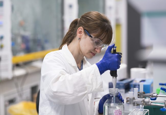
Proteins are the workhorses of biological systems but traditional methods for labelling them in living cells interfere with their function. Professor Ben Davis, Science Director for Next Generation Chemistry at the Franklin, and his team are developing new ways of altering proteins and other key biomolecules to study disease mechanisms at the atomic level.
“Many biomedical researchers are focusing on genetic approaches, but to understand how the genetic code conveys functions you have to study the workhorses, the proteins and the things they make – that is where the subtlety of biology lies,” Professor Davis says. Post-translational modifications and protein–protein interactions fine-tune protein function in a way that cannot be determined from genetic sequences alone. Yet, many researchers shy away from studying biology at the post-translational state because they can be much harder substrates to work with. “The genetic code may give us the ‘base’ recipe, but not necessarily the taste of the cake, as it were” he adds.
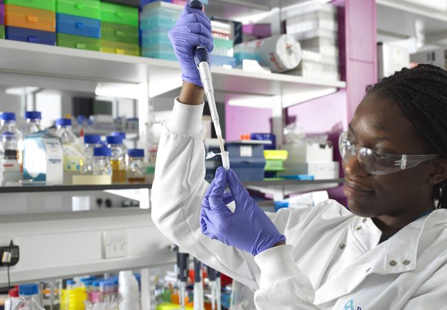
Adeline Poh, PhD student at the University of Oxford, is investigating biomarkers of neurodegeneration. Over the last 20 years, a growing number of studies have shown that neurofilament light chain (NfL) levels in the cerebrospinal fluid and blood are altered in neurological disorders and are correlated with disease characteristics1 . However, the relevance of NfL to disease is unclear.
Working in collaboration with researchers at The University of Oxford and Basel University Hospital, Ms Poh has generated radiolabelled NfL that is amenable to Positron Emission Tomography (PET) and introduced it into living mice to observe its behaviour in real time.
“NfL holds great promise as an early marker of neurodegeneration but to realise its potential value for diagnosis, prognosis and disease monitoring we want to understand if it contributes to pathogenicity at the atomic level,” she explains. “By inserting a tiny radiolabel that doesn’t perturb the native behaviour of NfL we can start to understand what happens to NfL in the brain and when it enters circulation.”
Ms Poh’s method can be used to track the movement of NfL from brain to serum, determine its preferred organ and to detect any changes that occur to the protein as it circulates through the body.
Choosing the right radioisotope and amino-acid residue to attach it to is key for this type of experiments. “When doing radiochemistry, time is against you,” says Ms Poh. Because the half-life of fluorine-18 is just under two hours, it requires a rapid and efficient method to position it on an amino acid residue that is not important for the protein’s native assembly so it won’t affect protein behaviour.
“The beauty of using fluorine as a radioisotope is that the length of a Carbon-Fluorine bond is somewhere between that of a Carbon-Hydrogen and a CarbonOxygen bond so it makes a very small perturbation to the structure,” Professor Davis explains. “Adeline’s work is some of the most powerful we have developed to unpick biological mechanisms in an artefact-free way.”
Professor Davis and Ms Poh are looking forward to applying the method to other biomarkers and proteins such as histones that regulate gene expression. They both agree that being able to study proteins involved in disease at the whole organism level has been a missing link in medicine and a lack of understanding of the molecular mechanisms underlying pathology is often at the root of drug discovery failures.
From tracking proteins to modifying their function
One of the Franklin’s goals is to chart this new world. Professor Davis’ work is integral to the Institute’s work on Atomic Pathology, which is trying to understand disease processes at the level of the bonds that are made and broken between atoms with the idea that it could lead to new physiological understanding and hence therapeutic interventions. His team is collaborating with multiple academic and industrial partners to create new possible modes of treatment such as logic-gated nanobodies, synthetic biologics, precise vaccines and covalent protein inhibitors.
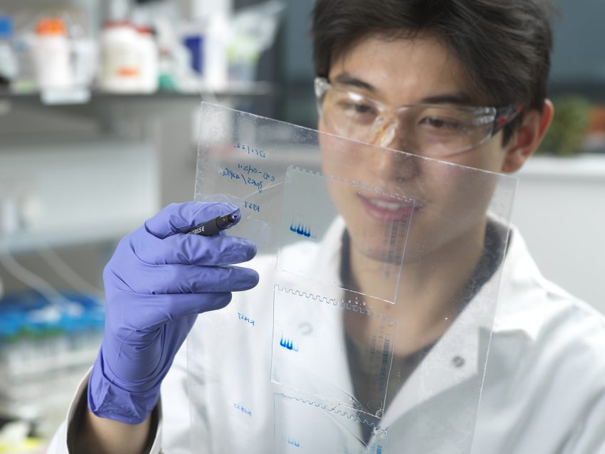
Previous work led by Professor Davis, developed in collaboration with Professor Veronique Gouverneur at the University of Oxford, has demonstrated how to specifically and selectively edit proteins using light2 . “Using these new protein editing methods, we can make protein surrogates with reactive side chains that are only activated by proximity,” Professor Davis says. Thus, they can control a protein’s reactivity so it can effectively bind or ‘trap’ disease-causing agents and remove them.
“This possibility of exploiting the protein–protein interface to drive therapy could lead to new biologics that overcome some of the limitations of current therapeutic antibodies, such as off-target effects and limited circulation half-life,” he says.
Other post-translational labelling methods that are being explored include precise manipulation of endogenous amino acids3,4 and inserting essentially abiotic elements, such as boron, into preselected sites to modify protein stability and function5 .
“Our approach is complementary to others; we hope to challenge the status quo in biology and biotechnology by developing new, essentially chemical, methods to edit organisms and hence probe and better understand their biology – this is a less common approach,” he explains. Close collaborations with pharmaceutical companies ensure that the methods being developed are relevant and creates the opportunities to change the treatment of disease.
“We want others to use our chemistry to further knowledge on biological mechanisms, challenge dogmas and drive disruptive innovation,”

1. Gaetani L, et al. Neurofilament light chain as a biomarker in neurological disorders. Journal of Neurology, Neurosurgery & Psychiatry 90, 870–881 (2019).
2. Josephson B, et al. Light-driven post-translational installation of reactive protein side chains. Nature 585, 530–537 (2020).
3. Imiołek M, et al. Residue-Selective Protein C-Formylation via Sequential DifluoroalkylationHydrolysis. ACS Cent. Sci., 7, 1, (2021).
4. Isenegger P G, et al. Posttranslational, site-directed photochemical fluorine editing of protein sidechains to probe residue oxidation state via 19F-nuclear magnetic resonance. Nature protocols, 18, 1543– 1562 (2023).
5. Mollner TA, et al. Post-translational insertion of boron in proteins to probe and modulate function. Nat Chem Biol 17, 1245–1261 (2021).