Remote testing of new imaging technology with the user community
A critical step in technology development is to test and refine new equipment with user groups, so their feedback shapes final designs and usability improvements.

Researchers at the Franklin have been working closely with Thermo Fisher Scientific through our Wellcome funded ‘Electrifying Life Sciences’ programme to develop the next generation of electron microscopes for imaging the smallest structures of cells in intact tissues, bring these tools to market and establish new methods and standards for the community to enable their wide use.
As one of the Franklin’s founding members, we have worked closely with Diamond Light Source from our inception. A commissioning call through the Diamond eBIC facility enabled first time community access to key components of our cryo-electron tomography pipeline.
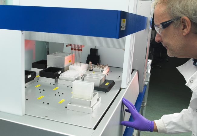
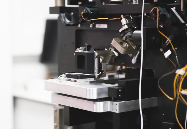
As part of a collaboration with ThermoFisher Scientific, teams from the Rosalind Franklin Institute and Diamond Light Source are offering academic researchers access to the latest generation of plasma focused ion beam (PFIB) scanning electron microscopes. Developed by ThermoFisher Scientific, the Helios Hydra 5 DualBeam’s unique capabilities are being tested and refined with the user community through a commissioning call funded by Wellcome and the Engineering and Physical Sciences Research Council.
Research from the Franklin’s Structural Biology Group shows the instrument has potential capabilities that extend beyond its intended use in sample preparation. With advanced volume imaging capabilities at cryogenic temperatures, it is possible to simultaneously image and slice through a sample, so users can find their area of interest and then conduct higher-resolution imaging on that region.
The beam used to mill the sample can be adapted to the sample’s specific make-up (e.g. silicon, metal or cell-based samples) thanks to the different plasma source ions, and the imaging is performed using an ultra-high resolution low acceleration voltage electron column. This flexibility, combined with the new technique proposed by the Franklin, allows for fast and easy sample processing, high-resolution surface imaging, and detailed analysis of cellular membranes, organelles and other structures. In the life sciences, this means the instrument can visualise how cellular membranes are organised and the distribution of organelles within a cell, offering potential applications in vaccine studies and infectious diseases research.
The commissioning call is part of a broader strategy to refine how the Franklin’s technologies and methods are disseminated, ensuring staff are prepared for user questions.
Dr Maud Dumoux, Associate Investigator in the Franklin’s Structural Biology Group, explains, “Rolling out new instrumentation at this early stage helps us to understand what we need to do to help users to adopt new technologies, and how these technologies can be designed and deployed in ways that make them user friendly. Although the Helios Hydra was designed to work with a specific workflow in mind, collaborating with users during our development work means we can further enhance its functionality based on real-world feedback.”
The call is integrated within Diamond’s peer-reviewed user programme and is open to academic users, as well as non-proprietary research from industry that will later be published. Users are required to have suitable samples and preliminary data demonstrating a need for cryo volume imaging in their research, and each is given 24 hours with the instrument. In return, users agree to provide feedback on their experience, workflow, results and areas for improvement, while also acknowledging support from both the Franklin and Diamond in their research publications.
While Diamond hosts the microscope and manages user selection and access, the Franklin’s Structural Biology Group integrates user feedback into their software, hardware and workflow development in partnership with ThermoFisher. This helps to optimise the imaging process and address issues raised by users before the instrument is made more widely available as an imaging tool.
“Projects like this help shape the design process for new instrumentation and provide scientific exemplars and use cases,” says Dr Daniel Clare, Principal Electron Microscopist at Diamond’s Electron Bio-Imaging Centre. “As a user facility, working together with the Franklin on this call allows us to introduce novel uses of an existing instrument to the user community as early as possible and helps us to make informed decisions about future technology investments.”
While there has been significant interest from the user community, accessing early-stage instrumentation in this way comes with challenges. “Although we’ve hosted this instrument for the past three years, this particular technique is quite complex to operate, so some of the output has been less than we would expect from other microscopes we have on-site,” says Diamond’s Dr Alistair Siebert, Principal Beamline Scientist, HeXI. “The technique we’re testing is relatively novel, speculative and early in its development life cycle, so it’s not quite ready to handle some of the samples that users have been interested in.”
Commissioning calls are common at Diamond, but this is only the Franklin’s second commissioning call and represents a new way to work with long-standing partners at Diamond in technology assessment.
While Diamond’s team are heavily involved in supporting users through the commissioning process, Dr Dumoux’s team remain at arm’s length from users and their projects to eliminate bias in their development work and maintain integrity.
“For the Franklin, this form of remote testing is a new way for us to quite quickly understand whether new technologies work and how users receive them before they’re fully mature and available,” explains Dr Dumoux. “We’re viewing this project as a test case to see if the commissioning call model is a good way to disseminate the Franklin’s technologies, or if other approaches are more suitable.”
More case studies
-
![]()
Franklin researchers demonstrate ‘significant potential’ of llama antibodies as potent Covid-19 treatment
-
![]()
Collaborating to shape and deliver the future of cryo-electron tomography
-
![]()
Using mass spectrometry imaging to understand metabolic mechanisms of disease
-
![]()
Peering into subcellular architecture at unprecedented resolution
-
![]()
BioCOP
-
![]()
University of Edinburgh