New stigmatic imaging prototype shows benefits of academic-industry partnering
The University of Oxford’s Department of Chemistry is presently home to one of our most exciting prototype technologies, the newly designed stigmatic imaging mass spectrometer.


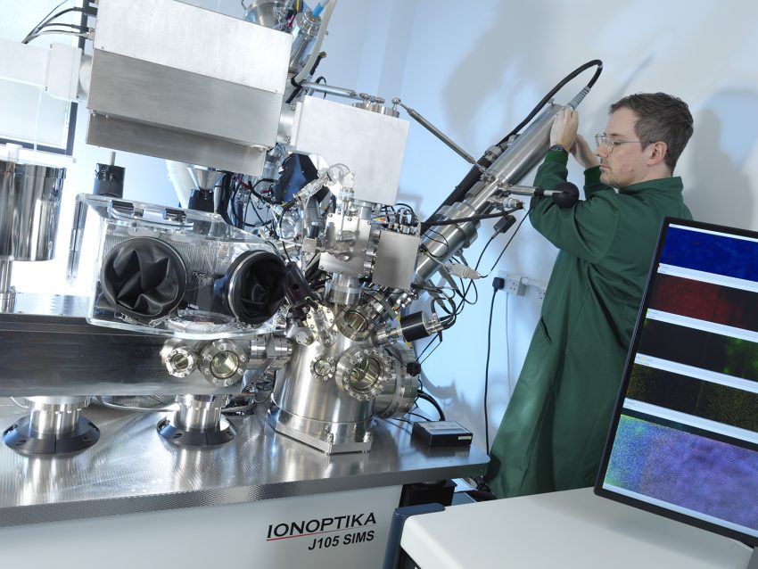
The instrument developed in collaboration with UK based Ionoptika enables a dramatic improvement in mass spectrometry imaging (MSI). The new instrument is capable of imaging the whole surface simultaneously, allowing more accurate imaging at faster speeds than was previously possible.

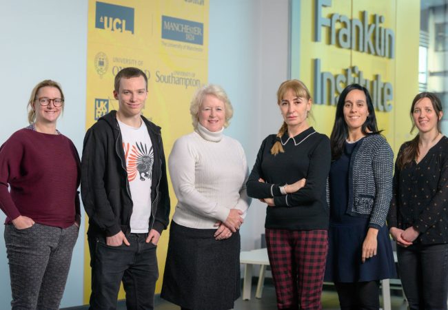
Scientists including Dr Felicia Green from our Biological Mass Spectrometry team have spent many months on the precise characterization and alignment of the primary ion beam.
Extraction optics and high-speed stigmatic detectors have now been fitted and the instrument is undergoing fine tuning and testing, so that all components can work to their optimum ability.
Whole-surface imaging
This instrument is particularly exciting because it has the potential to achieve a dramatic improvement in mass spectrometry imaging (MSI). Typically, MSI involves slow scanning across a surface, taking a mass spectrum at different points on the surface and gradually building up the image. But the new instrument will be capable of imaging the whole surface simultaneously, using the highest specification cameras that can operate as an array of position- and time-sensitive detectors, to record a mass spectrum for each pixel in the camera image. It will enable the most accurate images at much faster speed – and this would mean that analysing a standard tissue biopsy would take seconds, rather than hours or days, potentially allowing it to become part of routine analysis.
This world-class instrument design is a joint project between researchers at the Franklin, the University of Oxford, and the privately-owned ion beam technology company Ionoptika. Ionoptka’s scientists and engineers are strongly committed to the development of its technology for all scientific analysis applications, such as secondary ion mass spectrometry.
‘Our involvement grew and grew’
From the Franklin’s very earliest days, Ionoptika was closely involved in this particular project, as Kate McHardy, Ionoptika’s Sales Director, remembers.
‘When the Franklin was set up in 2018, we were invited to participate in various working groups that it was organising at the National Physical Laboratory, including those on different types of mass spectrometry imaging. We volunteered to be on the stigmatic imaging system working group, and initially our input was simply to advise on the instrument that was already planned – as well as to recondition an ion beam system that NPL were planning to lend to Oxford. But after lots of discussion and collaboration, our involvement grew – and we ended up designing and building the instrument too.’ In the first phase of the project, the bespoke SIMS instrument was built by Ionoptika at its headquarters near Southampton. It features a vacuum chamber, a stage and a sample handling system.
Separately, but as part of the same project, the team explored whether it might be possible to use a different ion beam technology on the new instrument, namely the use of Ionoptika’s gas cluster ion beam technology with water source, which is able to produce extremely good results from high molecular weight biological samples.
However, a feasibility study showed that this would be extremely challenging. The C60 ion beam gun was ultimately chosen for this prototype instrument. Dr Felicia Green spent time on site in Southampton helping with the build process. She said: ‘It was such a great experience; the team at Ionoptika were brilliant and very efficient. That was a really valuable period of exploration in finding out what was and was not possible.’
Next phase at Harwell
In April 2021 the prototype was moved to the Chemistry department at the University of Oxford, where the final parts of the instrument were fitted and tested in collaboration with their experts in time and position sensitive detectors, led by Professor Mark Brouard. Now the ion beam needs to be fully characterised, with the detector specifications calibrated and fine-tuned by the scientists there, and any additions made.
But this will not be the final iteration. Depending on results, information from this prototype will be used to build the next level instrument, destined for the Franklin’s Biological Mass Spectrometry instrumentation division at its hub in Harwell. Dr Green explained: ‘Our aim at Harwell is to build several instruments that can result in high throughput. This next spectrometer may be built on to a bigger instrument there, or could ultimately become a standalone unit capable of quick scans.’
Although Ionoptika’s commitment to the project is now officially complete, unofficially the collaboration remains in place, with regular virtual meetings with the projects’ in-situ scientists.
Kate McHardy says: ‘We are very much still interested, and hope to be involved when the next phase happens. It’s part of the way we are as a company to be involved from the start on exciting and challenging projects, because we have varied products that in effect can go in any direction.’
‘This rapid SIMS imaging has huge potential for the future and we’re watching closely to see what happens’.