Dorothy and Franklin
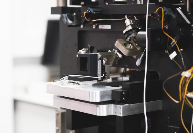
Ushering in a new era of cellular tomography. In the second half of 2021, the Rosalind Franklin Institute took delivery of its first Titan Krios cryo-electron microscope, named Dorothy, and the second generation of its plasma focused ion beam (pFIB), named Franklin.
As one of the Franklin’s founding members, we have worked closely with Diamond Light Source from our inception. A commissioning call through the Diamond eBIC facility enabled first time community access to key components of our cryo-electron tomography pipeline.

Researchers at the Franklin have been working closely with Thermo Fisher Scientific through our Wellcome funded ‘Electrifying Life Sciences’ programme to develop the next generation of electron microscopes for imaging the smallest structures of cells in intact tissues, bring these tools to market and establish new methods and standards for the community to enable their wide use.
Secured as part of the transformative £25m Electrifying Life Science grant from the Wellcome Trust, these cutting-edge technologies use new sample geometries to enable high-resolution, large-volume cellular tomography for the first time. The technology is being co-developed by scientists and engineers at the Rosalind Franklin Institute, Diamond Light Source, and manufacturers Thermo Fisher Scientific.
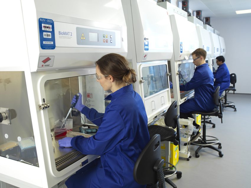
Large-volume cellular cryo-tomography – a method of obtaining 3D structures via cryogenic electron microscopy (cryo-EM) – has the potential to revolutionise our understanding of life, enabling us to characterise the structure of proteins at atomic resolution within the cell. Utilising the unique capabilities of these technologies will allow researchers to address more complex biological questions, including deepening our understanding of intracellular bacterial pathogen life cycles. Intracellular bacterial pathogens are becoming increasingly resistant to existing antibiotics, so the ability to observe the bacterial life cycle inside the human cell could be a critical first step in developing new drugs to combat this global threat.
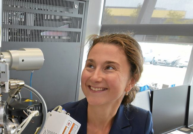
Critical innovations are required for this project to be successful – not only in the design of the instrument, but in the data collection and processing software used. Powerful data processing is needed to manage the volume and type of data produced, so close collaboration with the Franklin’s Artificial Intelligence and Informatics theme has proved essential. Dr Maud Dumoux, the Franklin’s technology lead for cryo-imaging, says: ‘This project is both challenging and exciting to be a part of – it has the potential to revolutionise how we see the cell. Improving our understanding of this basic building blocks of life could have a profound impact on how we create new drugs and understand how they work.’
Focused ion beam milling, as the name suggests, uses a stream of charged particles to prepare ultra-thin biological samples known as lamellae – a technique imported from materials science. In recent years, instruments using plasma beams of noble gases such as xenon have been developed to enable higherthroughput, higher-volume sample preparation. Used in combination with cryo-EM techniques, pFIB instruments like Franklin can streamline the previously cumbersome process of preparing and imaging small molecules inside the cell.
Dr Dumoux adds: ‘Until now, it has been very difficult to study the structure of molecules such as proteins at atomic resolution inside human cells. It involves lots of samples and multiple instruments, and can easily lead to the contamination, damage or loss of samples.
‘Our new workflow being developed at the Rosalind Franklin Institute will make this process much quicker and more efficient. By using pFIB to thin down the biological material to the required transparency, we then enable cryo-electron tomography of the internal regions of cells as part of a single pipeline. We have now demonstrated, for the first time, that these techniques can be used in combination for in-situ structural biology within cells – in this case, generating sub-nanometre-resolution data on particles called ribosomes.’
Plans are already in motion for a next-generation Titan Krios microscope – known as Hodgkin – which will be built as a collaboration between Thermo Fisher Scientific and the Rosalind Franklin Institute, and which is scheduled for delivery in 2024. There is already a clear vision for this machine, which will contain a special aberration corrector to allow collection of thicker biological samples. The knowledge gained from first-generation Dorothy will be crucial in informing the design of this beyond-state-of-the-art machine.
Steve Reyntjens, Product Marketing Director for Thermo Fisher Scientific, says: ‘Thermo Fisher is a company driven by the needs of scientists. It has been thrilling to work with the Rosalind Franklin Institute because they want to make breakthrough innovations, not just incremental changes – so working to meet their scientific needs has been a real challenge.’
“It has been thrilling to work with the Rosalind Franklin Institute because they want to make breakthrough innovations, not just incremental changes”.
The Electrifying Life Science grant was awarded to the Rosalind Franklin Institute and its partners the MRC Laboratory of Molecular Biology and Diamond Light Source to support the development of electron imaging physical sciences technologies with the capacity to transform how we see life. A globally unique resource for the UK, this suite of technologies will change by a factor of ten the accessibility and capability of cryo-EM in both tomography and single particle sub-fields.
Professor James Naismith, Director of the Rosalind Franklin Institute, adds: ‘The delivery of Dorothy in 2021 marked the start of the next stage of this project, and we are all excited to usher in the era of atomic cell biology. This type of high-risk, high-reward multidisciplinary project is what the UK government set up the Rosalind Franklin Institute to do. Electrifying Life Science will be a true factor-of-ten change in our ability to see and understand life, and we are committed to sharing our new advances with the widest community possible.’