Collaborating to shape and deliver the future of cryo-electron tomography
Researchers at The Rosalind Franklin Institute have been working closely with Thermo Fisher Scientific on a first-of-a-kind microscope that streamlines the generation of biological samples for cryo-electron tomography.

Researchers at the Franklin have been working closely with Thermo Fisher Scientific through our Wellcome funded ‘Electrifying Life Sciences’ programme to develop the next generation of electron microscopes for imaging the smallest structures of cells in intact tissues, bring these tools to market and establish new methods and standards for the community to enable their wide use.
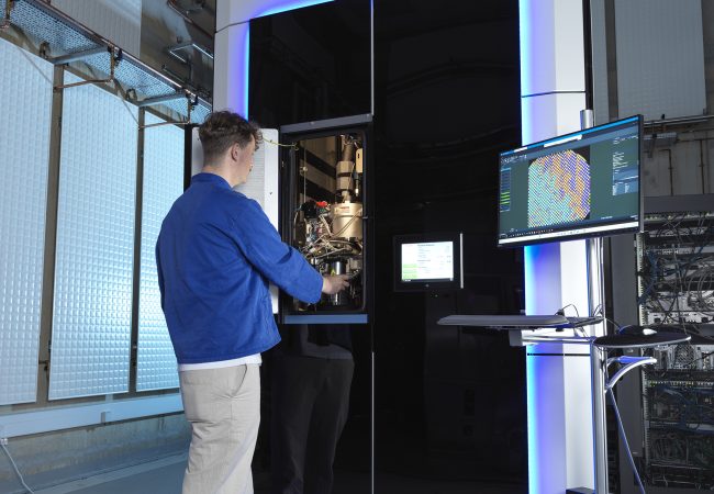
The Thermo Fisher ScientificTM ArctisTM Cryo-Plasma Focused Ion Beam (Cryo-PFIB) has been purposefully designed for thinning vitrified biological samples so they can be imaged in three dimensions using a transmission electron microscope (TEM).
“Until now, life sciences researchers doing cryo-electron tomography have worked with adapted instruments used for semiconductors or material science applications,” says Dr Steve Reyntjens, Senior Director of product marketing for electron microscopy at Thermo Fisher Scientific. “With Arctis they can produce large numbers of high-quality lamellae from vitrified cells to gain unprecedented molecular insights into biological processes.”
The collaboration between Thermo Fisher Scientific and researchers at the Franklin has been instrumental in getting Arctis Cryo-PFIB to market. “We started working on the concept of Arctis in 2019 as part of the Franklin’s mission to ‘electrify life sciences’,” Dr Reyntjens explains. “In the last 5 years, cryo-electron tomography has really gained traction as a powerful technique to study proteins in their cellular context, I’d say approximately half of the cryo-EM labs around the world have adopted cryo-electron tomography in some shape or form.”
Compared with traditional structural biology methods that analyse purified proteins or molecular complexes in isolation, in-cell cryo-electron tomography provides a much greater wealth of information. “By studying molecular structures in their close to natural-state, researchers can gather information about their location within cells and interacting partners, which is key to understand function.”
However, to realise the potential of cryo-electron tomography for life sciences several technical hurdles in the sample preparation process need to be overcome. The aim of the collaboration between Thermo Fisher Scientific and the Franklin was to bring about a stepchange in the methodology to further scientific discovery. “Over the two-year development period we thought long and hard about what is required to make Cryo-PFIB milling faster, easier and more successful,” Dr Reyntjens explains.
Following a continuous exchange of information throughout the design and build period, the first Arctis prototype was shipped to the Institute in mid-2021. By the end of the year, it was up and running and being used on a daily basis. “The timeline from defining the requirements to installing the working prototype at the Franklin was remarkable!” Reyntjens says.
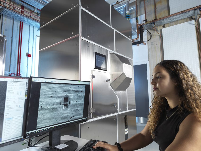
Arctis comprises a PFIB that provides multiple ion species (Xenon, Oxygen, Argon) for precision milling, as well as a scanning electron microscope and an integrated fluorescence light microscope to navigate the sample and target the region of interest.
Dr Reyntjens highlights two other important features for producing high-quality cryo-lamellae: the size of the vacuum chamber and connectivity with TEM, streamlining the cryo-electron tomography workflow. The small chamber helps maintain a high-quality vacuum and avoid ice contamination due to residual water molecules. In addition, the microscope’s autoloading system, facilitates the transfer of up to 12 grids at a time to the TEM, avoiding unnecessary manual handling and the risk of losing sample in the process.
Putting Arctis to the test
Dr Maud Dumoux, Technology Lead for cryo-imaging at the Franklin, and colleagues have been busy testing and reporting on the microscope’s performance. “As life sciences researchers with a broad range of expertise, from structural biology to pathology, we work to ensure that the equipment meets the needs of different communities and is easy to use,” she says.
“Any biologist studying molecular pathways will want to use Arctis”

Dr Dumoux has already published results from Arctis-produced lamellae1 . She is delighted with the microscope’s high throughput and full compatibility with TEM. “Before Arctis milling was very slow, impacting the workflow; with Arctis we can load up to 12 samples and run the equipment for days so we can finally increase the throughput.”
Now that the validity of hardware has been demonstrated, the Franklin and Thermo Fisher Scientific are working on improvements to the user interface and software. “We want to make it as accessible as possible, so a broader range of scientists will be able to use it,” Dr Dumoux says.
As Arctis microscopes are starting to be shipped all around the globe, Dr Reyntjens and Dr Dumoux are excited to see how they will be applied to life sciences research. “Any biologist studying molecular pathways will want to use Arctis,” Dr Dumoux says. “It is the machine everyone has been waiting for!”
1. Berger, C., Dumoux, M., Glen, T. et al. Plasma FIB milling for the determination of structures in situ. Nature Communications 14, 629 (2023). https://doi. o