BioCOP
The BioCOP: ‘pushing the boundaries’ of biological imaging across space and time.

A first of its kind piece of bespoke imaging technology arrived at the Franklin in April 2022, with the promise of revolutionising the way scientists observe living human cells.
The Biophotonic Correlative Optical Platform (BioCOP) allows researchers to study biological systems across multiple spatial and time scales simultaneously – at unprecedented resolution and sensitivity.
Once fully operational, The BioCOP will generate vital, detailed insights into human health and disease that were previously unattainable. The platform will tackle pressing research questions in human biology ranging from the structure of brain cells to the way our immune systems respond to threats from viruses or cancers.
The BioCOP is a partnership between the Franklin and the Kennedy Institute of Rheumatology at the University of Oxford, where the technology was housed during its design and assembly phases. The project sits within the Franklin’s Correlated Imaging theme, which aims to deepen our understanding of biological systems and processes by enabling information from multiple imaging techniques to be collected simultaneously. While the ‘correlation’ of data from different instruments is well established in fields such as astronomy, the concept has huge untapped potential in the life sciences.
The Franklin is the perfect place for this technology – it will benefit from on-site technical expertise in a range of areas, and there will be lots of opportunities for collaboration

According to project lead Professor Marco Fritzsche, an associate professor at the Kennedy and research fellow at the Franklin, the BioCOP will ‘push the boundaries’ of biological imaging. ‘To fully understand the biological processes happening in the human body during times of good health and illness,’ says Professor Fritzsche, ‘we need to be able to observe those processes across multiple space and time scales, in realistic and relevant tissue microenvironments.
‘The BioCOP allows us to do this by bringing together two cutting-edge imaging modalities at the same time. It’s a platform that pushes the boundaries of what we can currently do in biological imaging, and which fits in very neatly with the Franklin’s focus on making big technological leaps forward. The BioCOP will be up to 100 times faster, 100 times more sensitive, and allow ten times higher spatial resolution than previous technology.’
Replacing traditional imaging technologies
Specifically, the BioCOP system allows high-performance co-incidence and correlation imaging over multiple length and time scales, featuring a combination of fast high-throughput three-dimensional Lattice Light Sheet Microscopy (LLSM), super-resolution 3D Structural Illumination Microscopy (SIM), and minimally invasive long-term imaging within microfluidics at extended spatiotemporal resolution.
Quantitative correlative imaging of biological processes has become mission critical in the biomedical sciences. In recent years, state-of-the-art research has repeatedly demonstrated that the understanding of living systems demands technology with the capability of monitoring dynamic processes across a range of space and time scales, dissecting the function of living cells within their tissue microenvironments.
Novel developments such as super-resolution SIM and LLSM are transforming how living single cells and tissues can be studied, creating the expectation that these techniques will replace current imaging technologies such as confocal and widefield microscopy.
The therapeutics of the future
A key aspect of the BioCOP setup involves the creation of ‘organ-on-a-chip’ devices that mimic the environment inside the human body and enable the study of biological material – even down to individual cells – within tiny amounts of fluid. This will allow researchers to uncover the biophysical mechanisms taking place when, for example, immune cells interact with the antigens that provoke an inflammatory response to threats such as external pathogens. This has the potential to inform the diagnostic tools and therapeutics of the future.
Professor Fritzsche, whose Biophysical Immunology Laboratory probes at the interface between biology and physics in the context of the human immune system, highlights the ‘light touch’ nature of the BioCOP system as one of its chief benefits. He says: ‘In conventional imaging – particularly in fluorescence microscopy – the process of illuminating cells with lasers causes a phenomenon called phototoxicity. This can degrade the sample and influence the biology that’s taking place. The BioCOP’s superior speed and sensitivity allows us to carry out experiments much less invasively and destructively than before: we can observe cells over longer periods of time without producing those negative effects on the sample.’
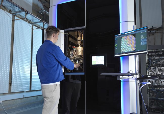
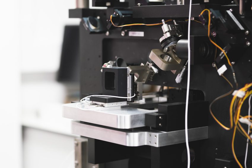
A one-metre-cubed ‘black box’ featuring a complex system of lasers and mirrors, the BioCOP is far removed from the traditional optical microscope design that many of us are familiar with. It will take, Professor Fritzsche estimates, up to two years of optimisation before BioCOP is ready to be used in experimental research.
He adds: ‘We have been very lucky in being able to recruit two excellent postdoctoral researchers, Dr Narain Karedla and Dr Anna Schepers, who will establish the BioCOP at the Franklin.
‘There has been a lot of excitement here about the arrival of the BioCOP and what it can do scientifically. The Franklin is the perfect place for this technology – it will benefit from on-site technical expertise in a range of areas, and there will be lots of opportunities for collaboration with researchers from other disciplines.’