Imaging neurodegeneration using mass spectrometry to understand the mechanisms of disease in the brain
Given the high complexity and heterogeneity of the cells in the brain, spatial information of molecular organisation is essential for the understanding of and insights into cellular processes.
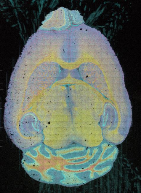
Secondary ion mass spectrometry (SIMS) and native ambient mass spectrometry (NAMS) imaging are increasingly recognised as a powerful technique for visualising molecular architectures in the fields of neurobiology and cell biology. SIMS provides the advantage of analysing multiple types of molecules, with cellular resolution, without the need for a prior knowledge and tagging. This project will use the unique instrumentation available at the Franklin for the generation of images with high spatial resolution and chemical specificity.