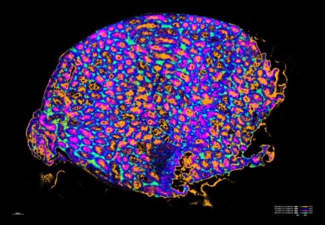
Mass spectrometry imaging
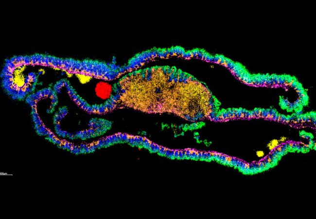
Subcellular Imaging
Next generation MS instrumentation will enable rapid molecular mapping of cells in tissue enabling elucidation of the chemistry behind biological mechanisms.
Biochemical Microscopy for imaging across Molecular Scales
Developing a transformative cryogenic 3D biochemical microscope, harnessing the power of high-resolution electron microscopy and mass spectrometry imaging
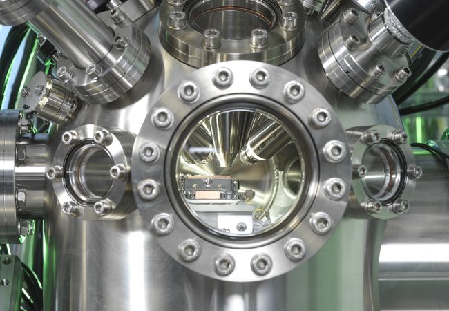
Chromatic Correction
Knoll, the first chromatic aberration-corrected electron microscope in the UK housed at the Franklin, will push the current resolution limits for biological samples by correcting energy variations in the electron beam.
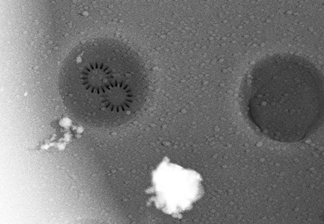
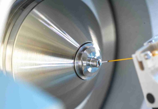
High Resolution imaging with secondary ion mass spectrometry (SIMS)
Secondary Ion Mass Spectrometry (SIMS) is a highly sensitive analytical technique offering detailed chemical composition analysis in 3D space with subcellular resolution.
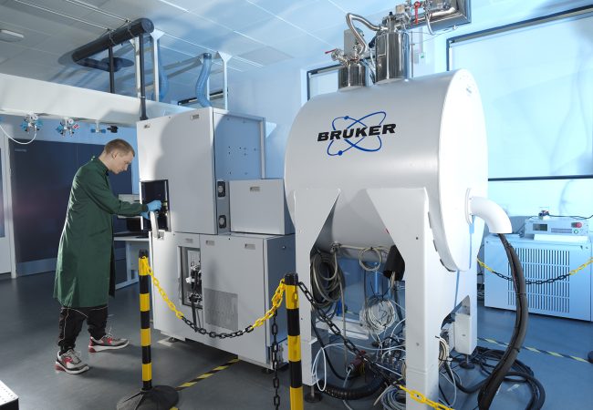
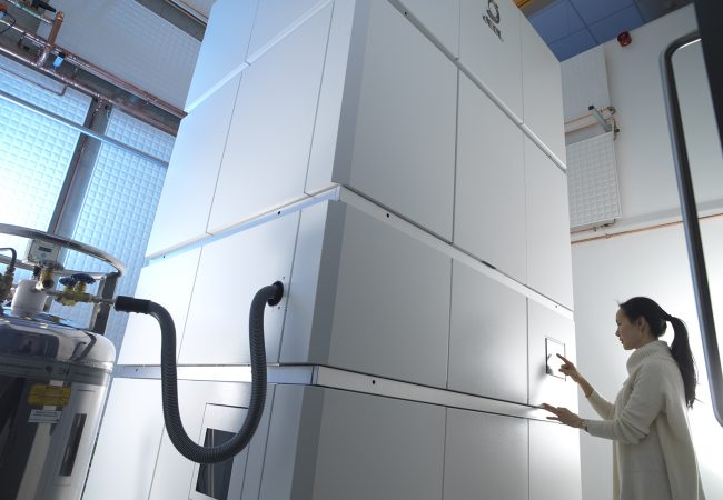
Trapped ion mobility (TIMS) time of flight (TOF) mass spectrometry
A cutting-edge commercial Bruker mass spectrometry (MS) instrument, coupling high sensitivity, high resolution, rapid time of flight (TOF) mass analysis to high resolution trapped ion mobility spectrometry (TIMS) enabling structural elucidation.
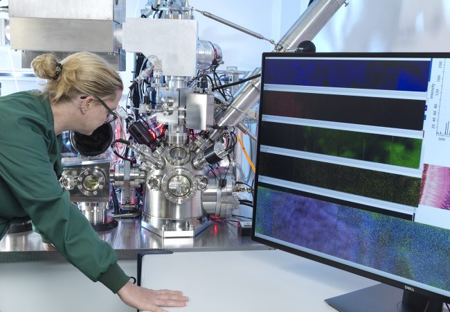
Native ambient mass spectrometry
Native ambient mass spectrometry (NAMS) combines spatial and structural biology by enabling untargeted label-free interrogation of proteins in their functional form directly from their physiological environment.
New tools for analysis of new drug modalities: Native ambient mass spectrometry of novel large molecule drugs
Native ambient mass spectrometry (NAMS) is an emerging field that enables label-free characterization of proteins and their complexes directly from their biological environment.
Dissecting the sugar-code in situ
Glycosylation – the covalent attachment of a sugar molecule to a protein – is the most abundant and indeed most complex post translational modification (PTM) in nature.