Cryo-EM
Experimental Automation
We integrate in situ structural biology, computer vision and research software engineering to develop imaging feedback–driven workflows for advanced microscopy. Our goals are to make high-resolution structural methods faster, more robust and accessible.
Structural Glycobiology
Complex carbohydrate molecules are essential for life. These diverse structures are found on every human cell and participate in a huge range of structural and signalling functions within cells, and between cells and the extracellular matrix.
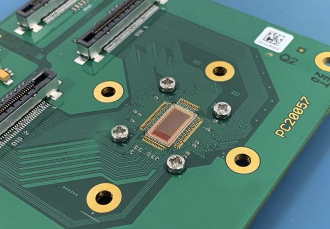
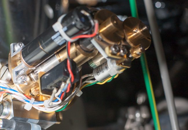
Electron Detector Development
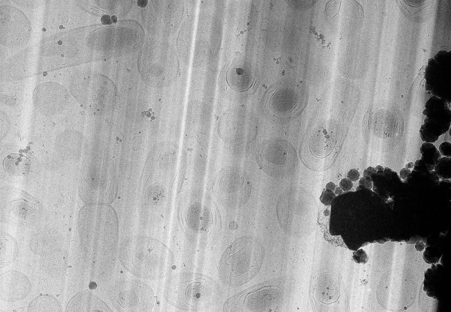
Atomic resolution imaging with electrons causes sample damage. The information per unit of damage is dependent on sample thickness and beam energy.
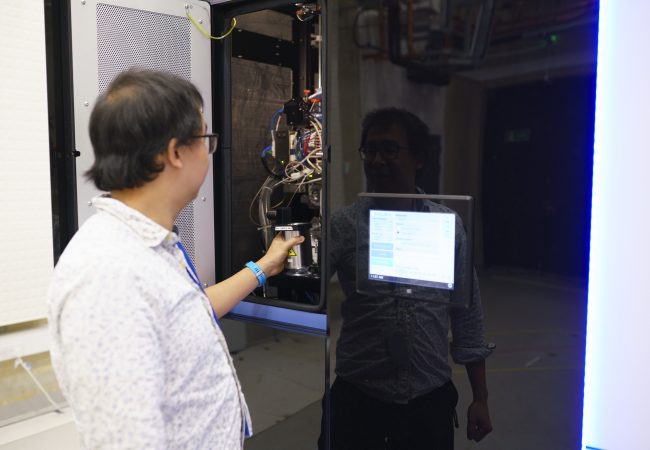
Aberration-corrected transmission electron microscope
Ruska is an aberration-corrected transmission electron microscope (TEM) used to explore novel methods to study radiation sensitive specimens such as biological materials that have been cryogenically preserved or encapsulated in liquid for dynamic observations.
Large Volume Tomography
High resolution large volume tomography with electron microscopy has the potential to transform our understanding of life, by linking the atomic and molecular structure of protein complexes in their biological context – the cell.
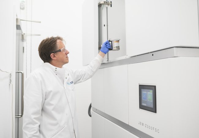
Cryogenic Electron Microscope (cryoARM)
Crewe is a first-generation cryogenic electron microscope based on JEOL’s atomic resolution microscope platform (ARM).
Chromatic Aberration-Corrected Electron Microscope
Knoll, the first chromatic aberration-corrected electron microscope in the UK, will push the current resolution limits for biological samples by correcting energy variations in the electron beam. This allows detailed imaging of thicker and liquid samples, benefiting biology by enabling…
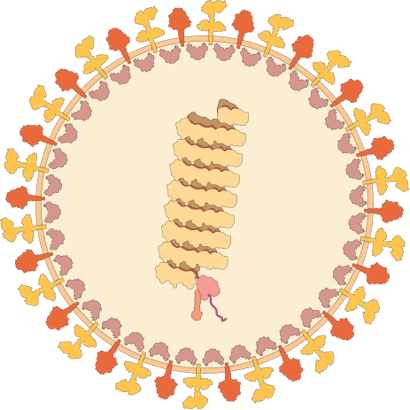
How Pathogens Interact with Human Cells
Our aim: To discover new ways of detecting, preventing and combatting human infectious diseases by discovering the mechanisms by which viruses and bacteria interact with human cells and tissues.
Multidimensional Imaging
Our aim: To develop new technologies to see the molecules of life in more detail including their dynamics and chemistry.
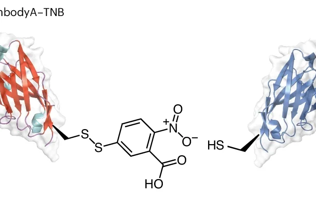
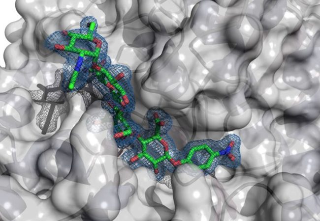
New bifunctional, bispecific nanobodies scaffold helps with small protein imaging
Researchers at the Rosalind Franklin Institute, University of Oxford, and Diamond Light Source have combined their expertise to create new scaffolding molecules to allow the electron microscopy imaging of small proteins (below 50 kDa). Using this method the team have…