Cryo-Ptycho-Tomography
Cryo-Ptycho-Tomography
Our recent work on cryo-EM ptychography (cryo-EP) demonstrated the application potential of the technique in characterising biological structure with dose-efficiency, signal-to-noise ratio, and large field-of-view. To further realise the potential of the technique, we are developing a new workflow of cryo-EM ptychographic tomography (cryo-EPT) and cryo-EM ptychographic subtomogram averaging and classification (cryoEPSTAC).
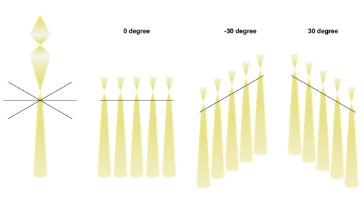
An advantage of the technique will be the flexibility of scanning probe control. Traditional cryo-ET suffers from defocus changes across large fields-of-view at higher tilting angles, while the defocus of a ptychographic scanning probe can be adjusted at a much more localised region. This would ensure the optimal acquisition geometry for the reconstruction.
Due to precise adjustments required for acquisitions, a significant proportion of microscope alignment, data acquisition and structure reconstruction will be automated using the live reconstruction results as a feedback control signal during acquisition. By combining accurate probe control for newly engineered acquisition geometries, we hope to enhance the output from tomographic methods using cryo-EP.

Project team members at The Franklin:
- Principal scientist: Chen Huang
- Judy Kim
- Emanuela Liberti
- The Franklin’s Artificial Intelligence and Informatics team